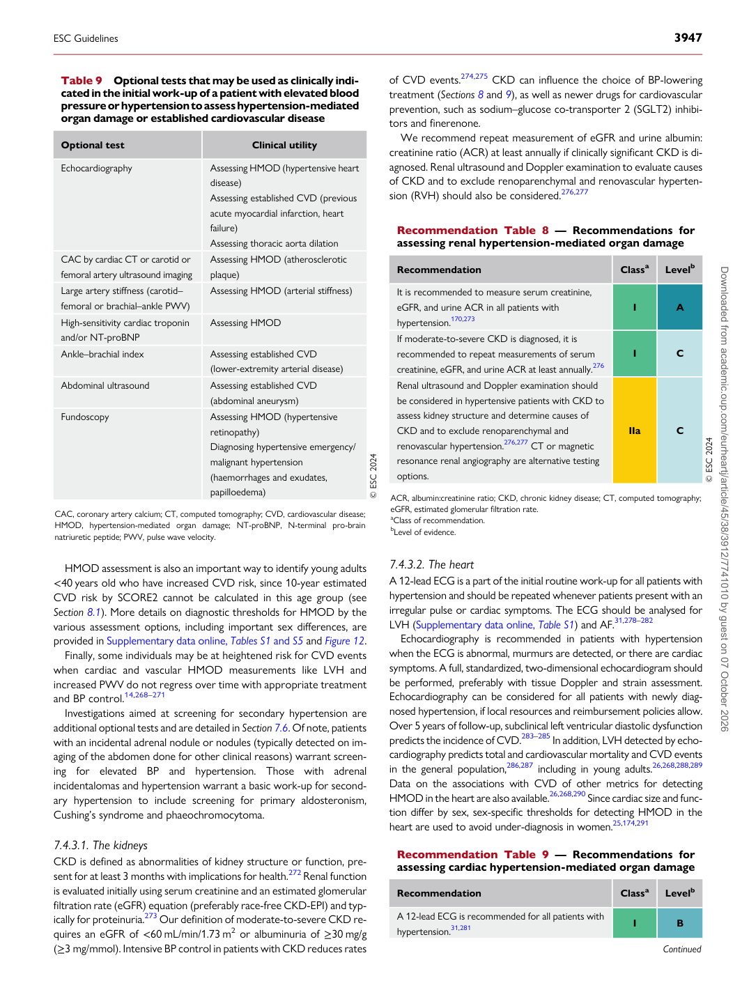
p.36 抽出テキスト
HMOD assessment is also an important way to identify young adults <40 years old who have increased CVD risk, since 10-year estimated CVD risk by SCORE2 cannot be calculated in this age group (see Section 8.1). More details on diagnostic thresholds for HMOD by the various assessment options, including important sex differences, are provided in Supplementary data online, Tables S1 and S5 and Figure 12. Finally, some individuals may be at heightened risk for CVD events when cardiac and vascular HMOD measurements like LVH and increased PWV do not regress over time with appropriate treatment and BP control.14,268–271 Investigations aimed at screening for secondary hypertension are additional optional tests and are detailed in Section 7.6. Of note, patients with an incidental adrenal nodule or nodules (typically detected on imaging of the abdomen done for other clinical reasons) warrant screening for elevated BP and hypertension. Those with adrenal incidentalomas and hypertension warrant a basic work-up for secondary hypertension to include screening for primary aldosteronism, Cushing’s syndrome and phaeochromocytoma.

7.4.3.1 The kidneys

CKD is defined as abnormalities of kidney structure or function, present for at least 3 months with implications for health.272 Renal function is evaluated initially using serum creatinine and an estimated glomerular filtration rate (eGFR) equation (preferably race-free CKD-EPI) and typically for proteinuria.273 Our definition of moderate-to-severe CKD requires an eGFR of <60 mL/min/1.73 m2 or albuminuria of ≥30 mg/g (≥3 mg/mmol). Intensive BP control in patients with CKD reduces rates of CVD events.274,275 CKD can influence the choice of BP-lowering treatment (Sections 8 and 9), as well as newer drugs for cardiovascular prevention, such as sodium–glucose co-transporter 2 (SGLT2) inhibitors and finerenone.

We recommend repeat measurement of eGFR and urine albumin: creatinine ratio (ACR) at least annually if clinically significant CKD is diagnosed. Renal ultrasound and Doppler examination to evaluate causes of CKD and to exclude renoparenchymal and renovascular hypertension (RVH) should also be considered.276,277

7.4.3.2 The heart

A 12-lead ECG is a part of the initial routine work-up for all patients with hypertension and should be repeated whenever patients present with an irregular pulse or cardiac symptoms. The ECG should be analysed for LVH (Supplementary data online, Table S1) and AF.31,278–282 Echocardiography is recommended in patients with hypertension when the ECG is abnormal, murmurs are detected, or there are cardiac symptoms. A full, standardized, two-dimensional echocardiogram should be performed, preferably with tissue Doppler and strain assessment. Echocardiography can be considered for all patients with newly diagnosed hypertension, if local resources and reimbursement policies allow. Over 5 years of follow-up, subclinical left ventricular diastolic dysfunction predicts the incidence of CVD.283–285 In addition, LVH detected by echocardiography predicts total and cardiovascular mortality and CVD events in the general population,286,287 including in young adults.26,268,288,289 Data on the associations with CVD of other metrics for detecting HMOD in the heart are also available.26,268,290 Since cardiac size and function differ by sex, sex-specific thresholds for detecting HMOD in the heart are used to avoid under-diagnosis in women.25,174,291

Table 9 Optional tests that may be used as clinically indicated in the initial work-up of a patient with elevated blood pressure or hypertension to assess hypertension-mediated organ damage or established cardiovascular disease

Optional test Clinical utility

Echocardiography Assessing HMOD (hypertensive heart

disease) Assessing established CVD (previous acute myocardial infarction, heart failure) Assessing thoracic aorta dilation

CAC by cardiac CT or carotid or femoral artery ultrasound imaging Assessing HMOD (atherosclerotic plaque) Large artery stiffness (carotid– femoral or brachial–ankle PWV) Assessing HMOD (arterial stiffness) High-sensitivity cardiac troponin and/or NT-proBNP Assessing HMOD Ankle–brachial index Assessing established CVD

(lower-extremity arterial disease)

Abdominal ultrasound Assessing established CVD

(abdominal aneurysm)

Fundoscopy Assessing HMOD (hypertensive

retinopathy) Diagnosing hypertensive emergency/ malignant hypertension (haemorrhages and exudates, papilloedema) © ESC 2024

CAC, coronary artery calcium; CT, computed tomography; CVD, cardiovascular disease; HMOD, hypertension-mediated organ damage; NT-proBNP, N-terminal pro-brain natriuretic peptide; PWV, pulse wave velocity. Recommendation Table 8 — Recommendations for assessing renal hypertension-mediated organ damage

Recommendation Classa Levelb

It is recommended to measure serum creatinine, eGFR, and urine ACR in all patients with hypertension.170,273 I A If moderate-to-severe CKD is diagnosed, it is recommended to repeat measurements of serum creatinine, eGFR, and urine ACR at least annually.276 I C Renal ultrasound and Doppler examination should be considered in hypertensive patients with CKD to assess kidney structure and determine causes of CKD and to exclude renoparenchymal and renovascular hypertension.276,277 CT or magnetic resonance renal angiography are alternative testing options. IIa C © ESC 2024

ACR, albumin:creatinine ratio; CKD, chronic kidney disease; CT, computed tomography; eGFR, estimated glomerular filtration rate. aClass of recommendation. bLevel of evidence.

Recommendation Table 9 — Recommendations for assessing cardiac hypertension-mediated organ damage

Recommendation Classa Levelb

A 12-lead ECG is recommended for all patients with hypertension.31,281 I B

Continued

ESC Guidelines                                                                                                                                                                                          3947

Downloaded from academic.oup.com/eurheartj/article/45/38/3912/7741010 by guest on 07 October 2026