p.81 抽出テキスト
6.4 Treatment of myocardial

revascularization failure One in five revascularized patients needs a repeat revascularization within the first 5 years after myocardial revascularization, with higher risk after PCI compared with CABG.1182 Revascularization failure can manifest either shortly after the initial procedure (within 30 days) or later on, and recurring symptoms may result from either restenosis of the treated coronary segment or the failure of bypass grafts,772 alongside the progression of underlying native CAD.1183,1184 Published evidence regarding diagnosis and management of myocardial revascularization failure has been summarized in the 2020 EAPCI (European Association of Percutaneous Cardiovascular Interventions) Expert Consensus Paper.1182 6.4.1 Percutaneous coronary intervention failure

Stent thrombosis and ISR are the most frequent reasons for PCI failure. Stent thrombosis occurs infrequently and is multifactorial. Anatomical and mechanical factors, as well as lack of adherence or hyporesponsiveness to antiplatelet treatment, are frequently the reasons behind this.1182,1185 The majority of patients with stent thrombosis present with ACS and should be treated according to the 2023 ESC Guidelines for the management of patients with acute coronary syndromes.65 Urgent ICA to confirm diagnosis and treatment is indicated. After restoration of coronary flow, intracoronary imaging to identify mechanical failure should be performed. Repeated DES implantation is indicated in case of stent fracture or collapse and residual edge dissections, while high-pressure non-compliant balloon dilation is indicated in case of stent under-expansion or malapposition. In-stent restenosis results as a response to vessel wall injury (neointimal hyperplasia) or neoatherosclerosis in the stented segment of the coronary artery. Although significantly less frequent than after bare-metal stent implantation, the incidence of clinical in-DES restenosis is up to 10% within the first 10 years after DES implantation1182 and remains the most frequent cause of PCI failure. The clinical presentation of ISR is mostly CCS, with 20% ACS, and the remaining asymptomatic. The indication to treat ISR is like that for native CAD. Radiological stent enhancement and intracoronary imaging are encouraged to determine the ISR mechanism. PCI treatment of ISR should be focused on the stenotic segment. Lesion preparation (ultra-high pressure balloon dilation, intravascular lithotripsy, rotation atherectomy) and correction of mechanical issues are required.1182 Thereafter, drug-coated balloon angioplasty or DES implantation is necessary.1186,1187 Drug-eluting balloon angioplasty and repeat stenting with DES were equally effective and safe in treating bare-metal ISR, but drug-coated balloon angioplasty was less effective than repeat paclitaxel DES implantation in treating DES ISR.1186 However, at 10-year follow-up there was no difference in clinical endpoints between drug-coated balloon angioplasty and DES implantation, whereas both were more effective than balloon angioplasty in preventing target-lesion revascularization.1187 Everolimus DES was associated with better long-term outcomes than drug-coated balloons.1188

6.4.2 Managing graft failure after coronary artery

bypass grafting A variety of reasons have the potential to adversely affect bypass graft patency.1189 These include technical (quality of graft material, surgical precision) and pathophysiological aspects (competitive flow, activity of the coagulation system, disease progression, etc.). Technical aspects and competitive flow are thought to influence early graft failure, while disease progression and graft degeneration affect long-term patency.1182,1189 The majority of graft occlusions are clinically silent.1189 If symptoms occur, prompt diagnostic workup (including ECG, assessment of biomarkers, and possibly repeat coronary angiography) is warranted to limit or prevent potential damage from graft occlusion.316 Acute CABG graft failure (<1 month after surgery) is observed in approximately 12% of grafts mostly due to technical problems.1190 Late failure of saphenous vein grafts occurs in up to 50% at 10 years, with vein graft occlusion rates in up to 27% within 1 year after surgery. 771,1191 The decision for optimal treatment (conservative, CABG revision/ redo CABG or PCI of the native vessel or of the failed graft) should be made individually considering haemodynamic stability, technical

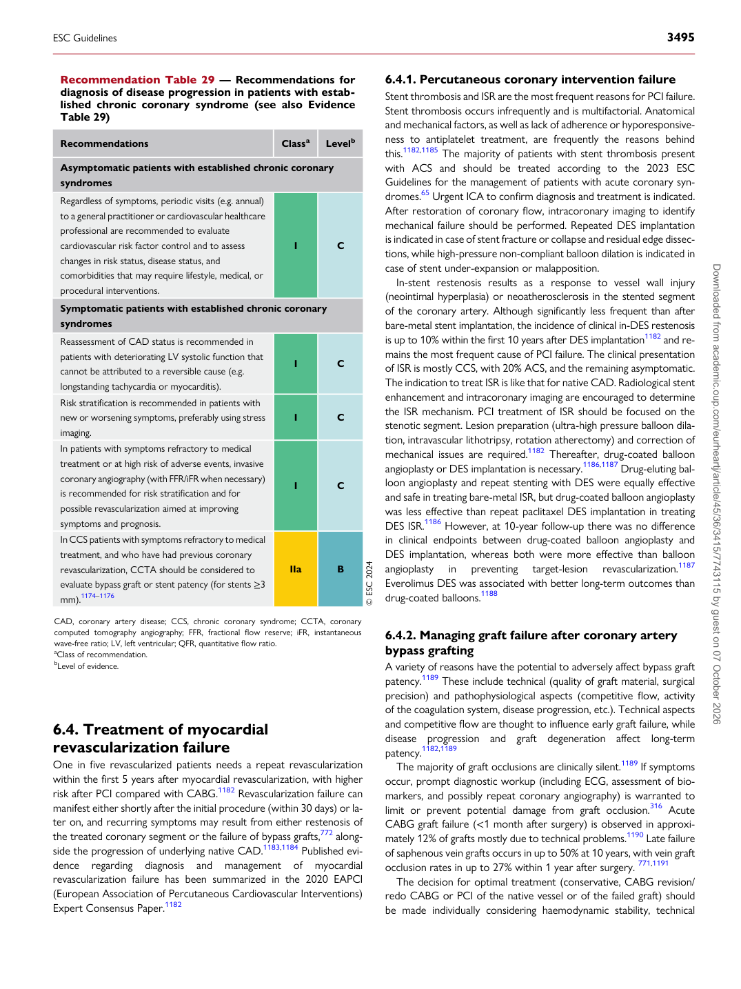
Recommendation Table 29 — Recommendations for diagnosis of disease progression in patients with established chronic coronary syndrome (see also Evidence Table 29)

Recommendations Classa Levelb

Asymptomatic patients with established chronic coronary syndromes Regardless of symptoms, periodic visits (e.g. annual) to a general practitioner or cardiovascular healthcare professional are recommended to evaluate cardiovascular risk factor control and to assess changes in risk status, disease status, and comorbidities that may require lifestyle, medical, or procedural interventions. I C Symptomatic patients with established chronic coronary syndromes Reassessment of CAD status is recommended in patients with deteriorating LV systolic function that cannot be attributed to a reversible cause (e.g. longstanding tachycardia or myocarditis). I C Risk stratification is recommended in patients with new or worsening symptoms, preferably using stress imaging. I C In patients with symptoms refractory to medical treatment or at high risk of adverse events, invasive coronary angiography (with FFR/iFR when necessary) is recommended for risk stratification and for possible revascularization aimed at improving symptoms and prognosis. I C In CCS patients with symptoms refractory to medical treatment, and who have had previous coronary revascularization, CCTA should be considered to evaluate bypass graft or stent patency (for stents ≥3 mm).1174–1176 IIa B © ESC 2024

CAD, coronary artery disease; CCS, chronic coronary syndrome; CCTA, coronary computed tomography angiography; FFR, fractional flow reserve; iFR, instantaneous wave-free ratio; LV, left ventricular; QFR, quantitative flow ratio. aClass of recommendation. bLevel of evidence.

ESC Guidelines                                                                                                                                                                                          3495

Downloaded from academic.oup.com/eurheartj/article/45/36/3415/7743115 by guest on 07 October 2026