p.35 抽出テキスト
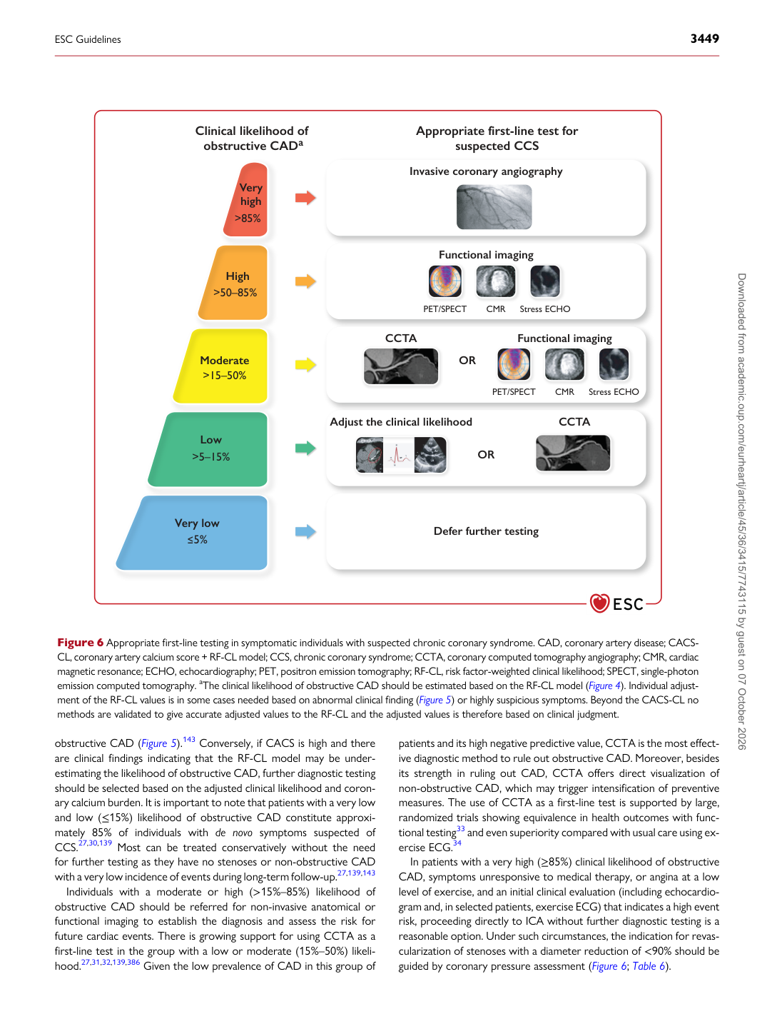
obstructive CAD (Figure 5).143 Conversely, if CACS is high and there are clinical findings indicating that the RF-CL model may be underestimating the likelihood of obstructive CAD, further diagnostic testing should be selected based on the adjusted clinical likelihood and coronary calcium burden. It is important to note that patients with a very low and low (≤15%) likelihood of obstructive CAD constitute approximately 85% of individuals with de novo symptoms suspected of CCS.27,30,139 Most can be treated conservatively without the need for further testing as they have no stenoses or non-obstructive CAD with a very low incidence of events during long-term follow-up.27,139,143 Individuals with a moderate or high (>15%–85%) likelihood of obstructive CAD should be referred for non-invasive anatomical or functional imaging to establish the diagnosis and assess the risk for future cardiac events. There is growing support for using CCTA as a first-line test in the group with a low or moderate (15%–50%) likelihood.27,31,32,139,386 Given the low prevalence of CAD in this group of patients and its high negative predictive value, CCTA is the most effective diagnostic method to rule out obstructive CAD. Moreover, besides its strength in ruling out CAD, CCTA offers direct visualization of non-obstructive CAD, which may trigger intensification of preventive measures. The use of CCTA as a first-line test is supported by large, randomized trials showing equivalence in health outcomes with functional testing33 and even superiority compared with usual care using exercise ECG.34 In patients with a very high (≥85%) clinical likelihood of obstructive CAD, symptoms unresponsive to medical therapy, or angina at a low level of exercise, and an initial clinical evaluation (including echocardiogram and, in selected patients, exercise ECG) that indicates a high event risk, proceeding directly to ICA without further diagnostic testing is a reasonable option. Under such circumstances, the indication for revascularization of stenoses with a diameter reduction of <90% should be guided by coronary pressure assessment (Figure 6; Table 6). Clinical likelihood of obstructive CADa Very high Appropriate first-line test for suspected CCS >85% High >50–85% Moderate >15–50% Low >5–15% Very low Invasive coronary angiography Functional imaging PET/SPECT CMR Stress ECHO OR Functional imaging CCTA CCTA Adjust the clinical likelihood OR Defer further testing 5% PET/SPECT CMR Stress ECHO Figure 6 Appropriate first-line testing in symptomatic individuals with suspected chronic coronary syndrome. CAD, coronary artery disease; CACS- CL, coronary artery calcium score + RF-CL model; CCS, chronic coronary syndrome; CCTA, coronary computed tomography angiography; CMR, cardiac magnetic resonance; ECHO, echocardiography; PET, positron emission tomography; RF-CL, risk factor-weighted clinical likelihood; SPECT, single-photon emission computed tomography. aThe clinical likelihood of obstructive CAD should be estimated based on the RF-CL model (Figure 4). Individual adjustment of the RF-CL values is in some cases needed based on abnormal clinical finding (Figure 5) or highly suspicious symptoms. Beyond the CACS-CL no methods are validated to give accurate adjusted values to the RF-CL and the adjusted values is therefore based on clinical judgment. ESC Guidelines 3449 Downloaded from academic.oup.com/eurheartj/article/45/36/3415/7743115 by guest on 07 October 2026