p.34 抽出テキスト
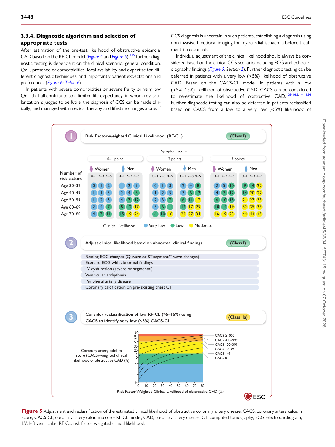
3.3.4. Diagnostic algorithm and selection of appropriate tests After estimation of the pre-test likelihood of obstructive epicardial CAD based on the RF-CL model (Figure 4 and Figure 5),139 further diagnostic testing is dependent on the clinical scenario, general condition, QoL, presence of comorbidities, local availability and expertise for different diagnostic techniques, and importantly patient expectations and preferences (Figure 6; Table 6). In patients with severe comorbidities or severe frailty or very low QoL that all contribute to a limited life expectancy, in whom revascularization is judged to be futile, the diagnosis of CCS can be made clinically, and managed with medical therapy and lifestyle changes alone. If CCS diagnosis is uncertain in such patients, establishing a diagnosis using non-invasive functional imaging for myocardial ischaemia before treatment is reasonable. Individual adjustment of the clinical likelihood should always be considered based on the clinical CCS scenario including ECG and echocardiography findings (Figure 5, Section 2). Further diagnostic testing can be deferred in patients with a very low (≤5%) likelihood of obstructive CAD. Based on the CACS-CL model, in patients with a low (>5%–15%) likelihood of obstructive CAD, CACS can be considered to re-estimate the likelihood of obstructive CAD.139,165,141,154 Further diagnostic testing can also be deferred in patients reclassified based on CACS from a low to a very low (<5%) likelihood of Risk Factor-weighted Clinical Likelihood (RF-CL) Symptom score Age 30–39 Very low Low Moderate Age 40–49 Age 50–59 Age 60–69 Age 70–80 Clinical likelihood: 0–1 point 2 points 3 points Number of risk factors 4 7 11 0 1 2 2 4 7 1 2 5 1 1 3 0–1 2–3 4–5 Women 15 19 24 1 2 5 8 12 17 4 7 12 2 4 8 0–1 2–3 4–5 Men 6 10 16 0 1 3 3 6 11 2 3 7 1 2 5 0–1 2–3 4–5 Women 22 27 34 2 4 8 12 17 25 6 11 17 3 6 12 0–1 2–3 4–5 Men 16 19 23 2 5 10 10 14 19 6 10 15 4 7 12 0–1 2–3 4–5 Women 44 44 45 9 14 22 32 35 39 21 27 33 14 20 27 0–1 2–3 4–5 Men Resting ECG changes (Q-wave or ST-segment/T-wave changes) LV dysfunction (severe or segmental) Exercise ECG with abnormal findings Ventricular arrhythmia Coronary calcification on pre-existing chest CT Peripheral artery disease 0 0 1 5 10 15 20 30 50 70 85 100 CACS 1000 CACS 400–999 CACS 100–399 CACS 10–99 CACS 1–9 CACS 0 Coronary artery calcium score (CACS)-weighted clinical likelihood of obstructive CAD (%) Risk Factor-Weighted Clinical Likelihood of obstructive CAD (%) 10 20 30 40 50 60 70 80 1 2 3 Consider reclassification of low RF-CL (>5–15%) using CACS to identify very low (5%) CACS-CL (Class I) (Class IIa) Adjust clinical likelihood based on abnormal clinical findings (Class I) Figure 5 Adjustment and reclassification of the estimated clinical likelihood of obstructive coronary artery disease. CACS, coronary artery calcium score; CACS-CL, coronary artery calcium score + RF-CL model; CAD, coronary artery disease; CT, computed tomography; ECG, electrocardiogram; LV, left ventricular; RF-CL, risk factor-weighted clinical likelihood. Downloaded from academic.oup.com/eurheartj/article/45/36/3415/7743115 by guest on 07 October 2026