p.32 抽出テキスト
In patients with suspected ANOCA/INOCA and an ICA/coronary pressure assessment disclosing no significant epicardial CAD, additional invasive investigations including index of microcirculatory resistance (IMR), CFR and, if necessary, invasive vasoreactivity testing using Ach (or ergonovine)36 as part of a complete ‘invasive coronary functional testing’ (ICFT) can be performed. Performing ICA is not exempt from potential complications. Given that femoral diagnostic catheterization has been associated with a 0.5%–2.0% composite rate of major complications, mainly bleeding requiring blood transfusions,327 radial access is now the standard access when possible. Radial access has been associated with reduced mortality and reduced major bleeding while allowing rapid ambulation.327 Still, the composite ICA rate of death, MI, or stroke through radial access is of the order of 0.1%–0.2%.327 The decision to perform ICA should balance benefits and risks, as well as potential therapeutic consequences, of the investigation that should be part of the process of shared clinical decision-making. Patients should be adequately informed of these aspects ahead of the procedure. 3.3.3.2 Functional assessment of epicardial stenosis severity to

guide coronary revascularization When non-invasive stress tests are inconclusive or not performed, identifying the artery responsible for ischaemia during ICA can be challenging, especially in cases with multivessel CAD or coronary stenoses of intermediate severity (typically around 40%–90% for non-left main stem stenoses or 40%–70% for left main stem stenoses by visual estimate). In such cases, recording wire-based intracoronary pressure during maximal hyperaemia to calculate FFR or at rest to measure iFR is recommended to improve risk assessment and clinical decision-making and to reduce clinical events.318–320 This has been confirmed by large clinical outcome studies such as FAME 1,308 FAME 2,49 DEFINE-FLAIR (Functional Lesion Assessment of Intermediate Stenosis to Guide Revascularisation),310 iFR-SWEDEHEART (Instantaneous Wave-free Ratio versus Fractional Flow Reserve in Patients with Stable Angina Pectoris or Acute Coronary Syndrome),311 R3F (French FFR Registry),313 and RIPCORD (Routine Pressure Wire Assessment Influence Management Strategy at Coronary Angiography for Diagnosis of Chest Pain trial).309 Haemodynamic relevance, as defined by FFR of ≤0.80, or iFR of ≤0.89, correlates poorly with diameter stenosis by visual assessment. In the PRIME-FFR [Insights From the POST-IT (Portuguese Study on the Evaluation of FFR-Guided Treatment of Coronary Disease) and R3F Integrated Multicenter Registries—Implementation of FFR (Fractional Flow Reserve) in Routine Practice]322 and FAME (Fractional Flow Reserve versus Angiography for Multivessel Evaluation) study,195 31% of the 40%–49% stenoses were haemodynamically significant while only 35% of the 50%–70% stenoses were haemodynamically relevant, and of the 71%–90% stenoses, 20% were not. Only an estimated diameter stenosis of >90% predicted haemodynamic relevance with high accuracy (96% correct classification). The discordance between angiographical and functional assessment of coronary stenosis severity varies with age, presence of CMD and lesionspecific factors.338,339 Lesions in the left main or proximal LAD are more likely to result in a significant FFR, as they supply a larger myocardial mass than those in smaller arteries. As a result, the optimal angiographic cut-off value for functionally non-significant stenosis is 43% for the left main and 55% for small vessels.339 This implies that the threshold for functional assessment for larger arteries should be set at 40% diameter stenosis. Large management studies showed that integration of FFR to ICA is associated with treatment reclassification in 30%–50% of cases in the R3F, POST-IT, RIPCORD, and DEFINE-REAL studies.309,313,340,341 Subsequently, many other non-hyperaemic pressure parameters were introduced [distal coronary pressure to aortic pressure ratio (Pd/Pa), diastolic pressure ratio (dPR), relative flow reserve (RFR)], with good correlation with FFR or iFR, but without available clinical outcome data. It is interesting to note that both separate and pooled analyses of the patients included in those studies reveal that ‘FFR/iFR-based reclassification’ does not have any significant effect on the number of patients recommended for revascularization.342 Meta-analyses of the 5-year outcome of patients managed with iFR and FFR as part of the randomized DEFINE-FLAIR and DEFINE- SWEDEHEART studies have reported a 2% absolute increase in all-cause mortality in those managed with iFR.343,344 This was not associated with any unplanned revascularization or non-fatal MI rate increase.343,344 Although it was initially hypothesized that this mortality excess could be related to a higher proportion of ‘inappropriate’ revascularization deferral with iFR compared with FFR (50% vs. 45%),343 it is reassuring that iFR-based deferral is as safe as FFR-based deferral up to 5 years.345 In patients with multivessel CAD, systematic FFR measurement of all epicardial vessels has been proposed to select appropriate therapy, but

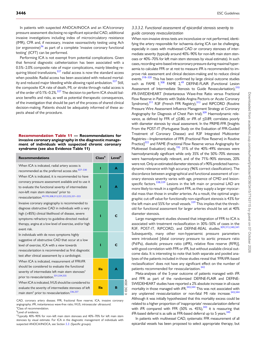
Recommendation Table 11 — Recommendations for invasive coronary angiography in the diagnostic management of individuals with suspected chronic coronary syndrome (see also Evidence Table 11)

Recommendations Classa Levelb

When ICA is indicated, radial artery access is recommended as the preferred access site.327–330 I A When ICA is indicated, it is recommended to have coronary pressure assessment available and to use it to evaluate the functional severity of intermediate non-left main stem stenosesc prior to revascularization.49,195,308,313,321,322,325,331–333 I A Invasive coronary angiography is recommended to diagnose obstructive CAD in individuals with a very high (>85%) clinical likelihood of disease, severe symptoms refractory to guideline-directed medical therapy, angina at a low level of exercise, and/or high event risk. I C In individuals with de novo symptoms highly suggestive of obstructive CAD that occur at a low level of exercise, ICA with a view towards revascularization is recommended as first diagnostic test after clinical assessment by a cardiologist. I C When ICA is indicated, measurement of FFR/iFR should be considered to evaluate the functional severity of intermediate left main stem stenosesc prior to revascularization.331,334,335 IIa A When ICA is indicated, IVUS should be considered to evaluate the severity of intermediate stenoses of left main stemc prior to revascularization.336,337 IIa B © ESC 2024

CAD, coronary artery disease; FFR, fractional flow reserve; ICA, invasive coronary angiography; iFR, instantaneous wave-free ratio; IVUS, intravascular ultrasound. aClass of recommendation. bLevel of evidence. cTypically 40%–90% for non–left main stem stenoses and 40%–70% for left main stem stenoses by visual estimate. For ICA in the diagnostic management of individuals with suspected ANOCA/INOCA, see Section 5.3. (Specific groups). Downloaded from academic.oup.com/eurheartj/article/45/36/3415/7743115 by guest on 07 October 2026