p.31 抽出テキスト
3.3.2.5 Non-invasive testing for microvascular dysfunction

Angina/ischaemia with non-obstructive coronary arteries (ANOCA/ INOCA) may be caused by transient and/or sustained impairments in the supply–demand of myocardial perfusion. Functional disorders leading to ANOCA/INOCA (e.g. MVA and VSA) are more common in women than in men.298,299 A recent meta-analysis reported an overall prevalence of MVA of 41% and VSA of 40% in selected patients without obstructive CAD.299 However, the true prevalence in unselected patient populations with suspected CCS remains unclear. Patients with ANOCA/INOCA have increased morbility/mortality,300,301 impaired quality of life (QoL), and weigh on health resource utilization. Early, accurate, and preferably non-invasive diagnosis is, therefore, of importance. The possibility of a microcirculatory origin of angina should be considered in individuals with symptoms suggestive of myocardial ischaemia and coronary arteries that are either normal or with non-obstructive lesions on CCTA or ICA. Several measurements that rely on quantifying blood flow through the coronary circulation are used to describe the function of the microvasculature to identify cases of MVA. Among the non-invasive imaging modalities, transthoracic Doppler echocardiography has been used as a non-invasive means to measure coronary blood flow but is limited to the assessment of the LAD artery and is affected by high inter- and intra-operator variability.302,303 Furthermore, this modality cannot distinguish between impairment of coronary flow caused by epicardial CAD or coronary microcirculatory dysfunction. A more direct and accurate microvascular function assessment is based on MBF measurement. This is commonly achieved by PET-CT myocardial perfusion imaging.299 PET allows for the quantification of MBF (expressed as millilitres per minute per gram of myocardium) and myocardial flow reserve (MFR). The latter reflects the magnitude of the increase in MBF that can be achieved by maximal coronary vasodilation conferred by vasodilators, such as adenosine or regadenoson. Since the microvasculature primarily determines vascular resistance, MFR measures the ability of the microvasculature to respond to a stimulus and therefore represents small vessel function. An MFR of less than 2.0 (2.5 for non-obstructive CAD) is often considered abnormal for PET.304 Of note, however, no definitive references are available across imaging modalities due to the moderate correlation among different MBF estimates.264 Recently, quantitative CMR has been proposed as an emerging technique for the assessment of microvascular dysfunction through MBF quantification but is currently limited to experienced centres.275

Quantitative myocardial perfusion can also be achieved by myocardial contrast echocardiography (MCE) through destruction–reperfusion imaging and analysis of the time–intensity curves from different regions of interest in the myocardium.231,233–235 Of note, MCE assesses capillary blood flow, and capillaries comprise 90% of the microvasculature. Measuring MBF at rest and during hyperaemia allows calculation of MBF reserve, which is associated with severity of coronary stenoses in patients with stable angina. In a meta-analysis, MBF reserve had high accuracy for predicting flow-limiting CAD.231 However, in the absence of obstructive CAD, reduced MBF reserve by MCE depicts microcirculatory abnormalities. Transthoracic Doppler evaluation of the LAD artery is also used to assess coronary flow reserve (CFR) during stress hyperaemia and has prognostic value.238,255,305,306 In contrast, the diagnosis of VSA ideally relies on the results of provocation tests in the catheterization laboratory through selective intracoronary acetylcholine (Ach) infusion (see Section 5.2.5.2). It is important to note that there is only a modest correlation between the values of MBF reserve measured by different techniques and modalities.269,305,307

3.3.3 Invasive tests

Invasive coronary angiography has undergone significant advancements over time. It is no longer just an angiographic technique that provides anatomical information about the presence of coronary atherosclerosis and luminal obstructions of the epicardial coronary arteries. It can also determine the functional consequences of these obstructions on coronary blood flow [FFR and instantaneous wave-free ratio (iFR)] by direct measurement of the coronary BP49,308–311 or by calculating the coronary pressure drop across a stenosis based on two or more angiographic projections.312 Furthermore, new technologies allow measurement of CFR and microvascular resistance, and protocols have been introduced for testing the presence of coronary vasospasm.36,39

3.3.3.1 Invasive coronary angiography

Invasive coronary angiography with available coronary pressure assessment49,308–311,313 is indicated in patients with a very high (>85%) clinical likelihood of obstructive CAD,1 in particular those with severe symptoms refractory to antianginal treatment, or characteristic angina or dyspnoea at a low level of exercise1,47 or left ventricle dysfunction suggesting extensive obstructive CAD.47,182,314,315 Invasive coronary angiography/coronary pressure assessment is also indicated if non-invasive assessment suggests high event risk—e.g. CCTA shows ≥50% left main stenosis, or ≥70% proximal LAD stenosis with single or two-vessel CAD, or ≥70% proximal three-vessel CAD56,182,316,317—or when any stress test shows moderate to severe inducible ischaemia316 or when symptoms are highly suggestive for obstructive CAD. In all the above situations, ICA/coronary pressure assessment is performed for additional risk stratification318–320 and to determine a potential revascularization approach (see Section 4.4).49,308,309,313 Invasive coronary angiography/coronary pressure assessment may also be indicated to confirm or exclude the diagnosis of obstructive CAD in patients with uncertain results on non-invasive testing.316 Given the frequent mismatch between the angiographic and haemodynamic severities of coronary stenoses, coronary pressure assessment should be readily available to complement ICA investigation for clinical decision-making.321–326

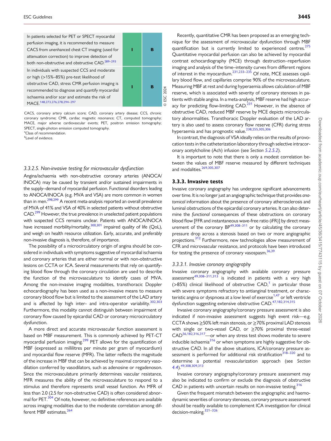
In patients selected for PET or SPECT myocardial perfusion imaging, it is recommended to measure CACS from unenhanced chest CT imaging (used for attenuation correction) to improve detection of both non-obstructive and obstructive CAD.289–293 I B In individuals with suspected CCS and moderate or high (>15%–85%) pre-test likelihood of obstructive CAD, stress CMR perfusion imaging is recommended to diagnose and quantify myocardial ischaemia and/or scar and estimate the risk of MACE.148,273,276,278,294–297 I B © ESC 2024

CACS, coronary artery calcium score; CAD, coronary artery disease; CCS, chronic coronary syndrome; CMR, cardiac magnetic resonance; CT, computed tomography; MACE, major adverse cardiovascular events; PET, positron emission tomography; SPECT, single-photon emission computed tomography. aClass of recommendation. bLevel of evidence.

ESC Guidelines                                                                                                                                                                                          3445

Downloaded from academic.oup.com/eurheartj/article/45/36/3415/7743115 by guest on 07 October 2026