p.30 抽出テキスト
However, non-obstructive coronary atherosclerosis not linked with ischaemia remains undetected by functional testing in general. If available, assessment of myocardial perfusion using SPECT is recommended in patients with suspected CCS with moderate or high pre-test likelihood of obstructive CAD (15%–85%) or known CCS. Importantly, if non-contrast-enhanced CT for attenuation correction is acquired, this allows for additional CAC scoring, providing important information for risk stratification even in the absence of flow-limiting coronary lesions.

3.3.2.3 Positron emission tomography-computed tomography

Similarly to myocardial perfusion SPECT imaging, PET also relies on radiopharmaceuticals. Contrary to SPECT, however, the radionuclides commonly used (i.e. 13N-ammonia, 15O-water, and 82Rubidium) are short-lived, with half-lives in the range of minutes, requiring production of these radionuclides ad hoc for every investigation. As attenuation correction is mandatory, PET is routinely performed in combination with non-contrast-enhanced CT. Scans are performed during both rest and infusion of pharmacological stressors (e.g. dobutamine) or vasodilators (e.g. dipyridamole, adenosine, or regadenoson). While myocardial perfusion PET-CT produces retention images depicting relative differences in regional MBF similar to those from SPECT —albeit with superior image quality and at much lower radiation dose exposure—the unique strength of PET-CT imaging is its ability to provide robust absolute quantitative measures of MBF. Measuring MBF with cardiac PET does not increase radiation or imaging time. Several measurements of MBF can be routinely obtained, including MBF during hyperaemia, MBF at rest, the MBF reserve, and the relative MBF reserve, and confer added diagnostic and prognostic value beyond relative perfusion assessment.262,263 Quantitative measures of MBF offer the ability to assess individuals with known or suspected diffusely impaired MBF, e.g. with multivessel CAD, or microvascular dysfunction.45,264 In general, PET-CT myocardial perfusion imaging is associated with high accuracy for detecting flow-limiting coronary lesions,148,258,265 and has been shown to provide prognostic information.223,262,263 In several head-to-head comparisons, PET-CT myocardial perfusion imaging outperformed other functional imaging modalities.257,266–269 However, whether the superiority in diagnostic accuracy leads to improved clinical effectiveness and post-test management remains to be elucidated.270 In a large retrospective study, a low MBF reserve measured by PET independently predicted mortality and helped identify patients with a survival benefit from early revascularization with PCI or coronary artery bypass grafting (CABG) beyond the extent of myocardial ischaemia.271 Limitations of PET-CT arise from its limited availability compared with other imaging modalities. Furthermore, methodological heterogeneity exists, particularly regarding thresholds for abnormality of quantitative measurements. Finally, physical exercise is challenging to perform. If available, assessment of myocardial perfusion using PET-CT is particularly recommended in obese patients (due to the high photon energy), in young patients (due to the low radiation dose exposure), and in those with known or suspected diffusely impaired MBF, e.g. those with multivessel CAD or microvascular dysfunction.264 Notably, the mandatory non-contrast-enhanced CT for attenuation correction allows for additional CAC scoring, providing essential information for risk stratification even in the absence of flow-limiting coronary lesions.

3.3.2.4 Cardiac magnetic resonance imaging

Aside from providing highly accurate and reproducible assessments of overall cardiac anatomy, cardiac volumes, function, and tissue characterization, CMR also offers the ability to assess myocardial perfusion, which relies on the first-pass myocardial perfusion of gadolinium-based contrast agents.

Recently, CMR methods using various parameters for quantitative MBF assessment have been introduced. However, the diagnostic performance of these parameters varies extensively among studies, and standardized protocols and software are lacking.272 Therefore, visual assessment of perfusion defects is currently used in clinical practice. Myocardial perfusion imaging by stress CMR combines high spatial resolution with the absence of ionizing radiation. This has been shown to provide high diagnostic accuracy in detecting flow-limiting coronary lesions,148,257,258 prognostic value,223,273–275 and improving patient management.178,276 Pharmacological vasodilators (e.g. adenosine or regadenoson) or stressors (e.g. dobutamine) are commonly applied, as physical exercise is challenging to perform. In conjunction with a dobutamine infusion, wall motion abnormalities induced by ischaemia can also be detected.277 Of note, and as for all non-invasive imaging modalities used for assessing myocardial perfusion, incorporating all available imaging and non-imaging information as part of an integrative approach is mandatory. For CMR, a multiparametric protocol, including LV function and assessment of LGE along with myocardial perfusion, increases the ability to rule in or rule out obstructive CAD in suspected CCS.278 Coronary magnetic resonance angiography allows non-invasive visualization of the coronary arteries.279 However, CMR angiography remains primarily a research tool due to limitations arising from long imaging times, low spatial resolution, and operator dependency. General limitations of CMR for myocardial perfusion arise from its limited availability, the claustrophobia experienced by patients, duration of image acquisition,280 and possible contraindications to CMR [e.g. nonconditional pacemakers and implantable cardioverter defibrillators (ICDs)] or to gadolinium-based contrast agents (e.g. renal failure due to the potential risk of nephrogenic systemic fibrosis). Finally, and contrary to SPECT/CT or PET-CT, stress CMR does not currently provide information on presence or absence of coronary calcifications. If available, and if no contraindications are met, stress CMR is recommended as an option in patients with suspected CCS with moderate or high (>15%–85%) pre-test likelihood of obstructive CAD or known CCS, particularly if additional information on cardiac function and tissue characterization is warranted.

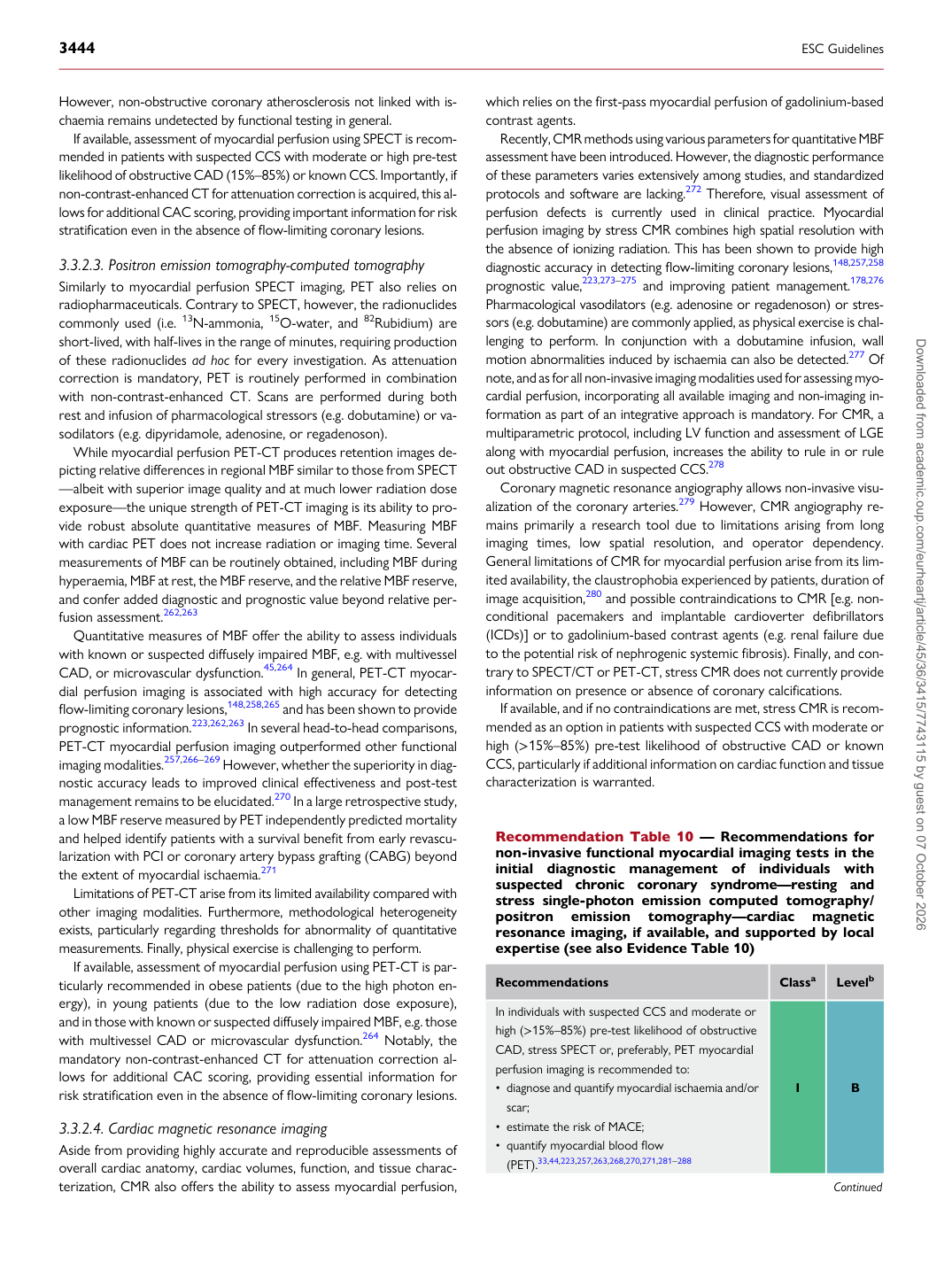
Recommendation Table 10 — Recommendations for non-invasive functional myocardial imaging tests in the initial diagnostic management of individuals with suspected chronic coronary syndrome—resting and stress single-photon emission computed tomography/ positron emission tomography—cardiac magnetic resonance imaging, if available, and supported by local expertise (see also Evidence Table 10)

Recommendations Classa Levelb

In individuals with suspected CCS and moderate or high (>15%–85%) pre-test likelihood of obstructive CAD, stress SPECT or, preferably, PET myocardial perfusion imaging is recommended to: • diagnose and quantify myocardial ischaemia and/or

scar; • estimate the risk of MACE; • quantify myocardial blood flow

(PET).33,44,223,257,263,268,270,271,281–288 I B

Continued Downloaded from academic.oup.com/eurheartj/article/45/36/3415/7743115 by guest on 07 October 2026