p.3 抽出テキスト
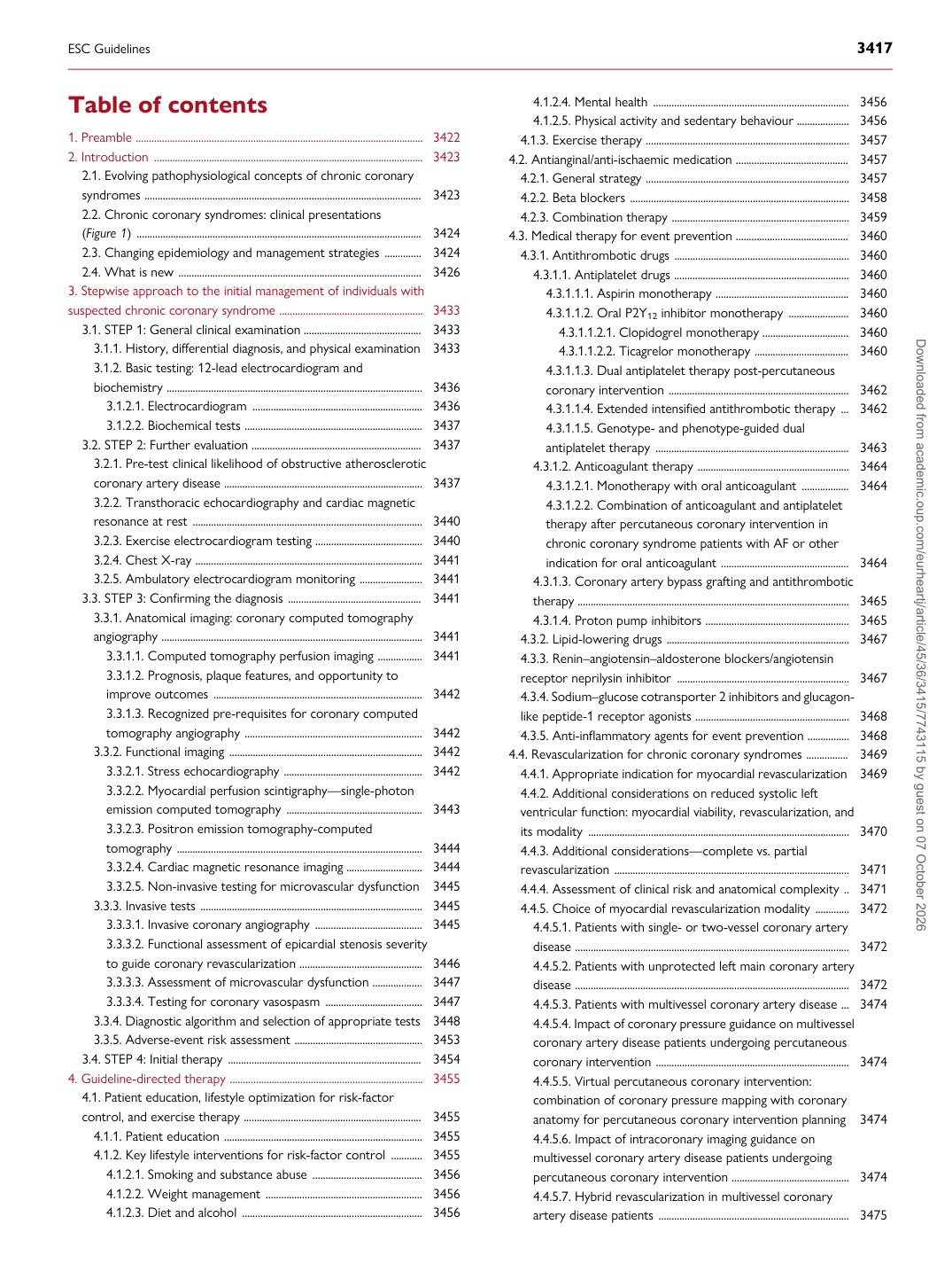
Table of contents 1. Preamble .............................................................................................................. 3422 2. Introduction ....................................................................................................... 3423 2.1. Evolving pathophysiological concepts of chronic coronary syndromes .......................................................................................................... 3423 2.2. Chronic coronary syndromes: clinical presentations (Figure 1) ............................................................................................................. 3424 2.3. Changing epidemiology and management strategies .............. 3424 2.4. What is new ............................................................................................. 3426 3. Stepwise approach to the initial management of individuals with suspected chronic coronary syndrome ....................................................... 3433

3.1. STEP 1: General clinical examination ............................................. 3433 3.1.1. History, differential diagnosis, and physical examination 3433 3.1.2. Basic testing: 12-lead electrocardiogram and biochemistry .................................................................................................. 3436 3.1.2.1. Electrocardiogram ................................................................. 3436 3.1.2.2. Biochemical tests .................................................................... 3437 3.2. STEP 2: Further evaluation ................................................................. 3437 3.2.1. Pre-test clinical likelihood of obstructive atherosclerotic coronary artery disease ............................................................................ 3437 3.2.2. Transthoracic echocardiography and cardiac magnetic resonance at rest ........................................................................................ 3440 3.2.3. Exercise electrocardiogram testing ......................................... 3440 3.2.4. Chest X-ray ....................................................................................... 3441 3.2.5. Ambulatory electrocardiogram monitoring ........................ 3441 3.3. STEP 3: Confirming the diagnosis ................................................... 3441 3.3.1. Anatomical imaging: coronary computed tomography angiography .................................................................................................... 3441 3.3.1.1. Computed tomography perfusion imaging ................. 3441 3.3.1.2. Prognosis, plaque features, and opportunity to improve outcomes ................................................................................ 3442 3.3.1.3. Recognized pre-requisites for coronary computed tomography angiography .................................................................... 3442 3.3.2. Functional imaging .......................................................................... 3442 3.3.2.1. Stress echocardiography ..................................................... 3442 3.3.2.2. Myocardial perfusion scintigraphy—single-photon emission computed tomography .................................................... 3443 3.3.2.3. Positron emission tomography-computed tomography .............................................................................................. 3444 3.3.2.4. Cardiac magnetic resonance imaging ............................. 3444 3.3.2.5. Non-invasive testing for microvascular dysfunction 3445 3.3.3. Invasive tests ..................................................................................... 3445 3.3.3.1. Invasive coronary angiography ......................................... 3445 3.3.3.2. Functional assessment of epicardial stenosis severity to guide coronary revascularization ............................................... 3446 3.3.3.3. Assessment of microvascular dysfunction ................... 3447 3.3.3.4. Testing for coronary vasospasm ..................................... 3447 3.3.4. Diagnostic algorithm and selection of appropriate tests 3448 3.3.5. Adverse-event risk assessment ................................................. 3453 3.4. STEP 4: Initial therapy .......................................................................... 3454 4. Guideline-directed therapy .......................................................................... 3455 4.1. Patient education, lifestyle optimization for risk-factor control, and exercise therapy .................................................................... 3455 4.1.1. Patient education ............................................................................ 3455 4.1.2. Key lifestyle interventions for risk-factor control ............ 3455 4.1.2.1. Smoking and substance abuse .......................................... 3456 4.1.2.2. Weight management ............................................................ 3456 4.1.2.3. Diet and alcohol ..................................................................... 3456 4.1.2.4. Mental health ........................................................................... 3456 4.1.2.5. Physical activity and sedentary behaviour .................... 3456 4.1.3. Exercise therapy .............................................................................. 3457 4.2. Antianginal/anti-ischaemic medication ........................................... 3457 4.2.1. General strategy .............................................................................. 3457 4.2.2. Beta blockers .................................................................................... 3458 4.2.3. Combination therapy .................................................................... 3459 4.3. Medical therapy for event prevention ........................................... 3460 4.3.1. Antithrombotic drugs ................................................................... 3460 4.3.1.1. Antiplatelet drugs ................................................................... 3460 4.3.1.1.1. Aspirin monotherapy ................................................... 3460 4.3.1.1.2. Oral P2Y12 inhibitor monotherapy ....................... 3460 4.3.1.1.2.1. Clopidogrel monotherapy ................................. 3460 4.3.1.1.2.2. Ticagrelor monotherapy .................................... 3460 4.3.1.1.3. Dual antiplatelet therapy post-percutaneous coronary intervention ..................................................................... 3462 4.3.1.1.4. Extended intensified antithrombotic therapy ... 3462 4.3.1.1.5. Genotype- and phenotype-guided dual antiplatelet therapy .......................................................................... 3463 4.3.1.2. Anticoagulant therapy .......................................................... 3464 4.3.1.2.1. Monotherapy with oral anticoagulant .................. 3464 4.3.1.2.2. Combination of anticoagulant and antiplatelet therapy after percutaneous coronary intervention in chronic coronary syndrome patients with AF or other indication for oral anticoagulant ................................................. 3464 4.3.1.3. Coronary artery bypass grafting and antithrombotic therapy ........................................................................................................ 3465 4.3.1.4. Proton pump inhibitors ....................................................... 3465 4.3.2. Lipid-lowering drugs ...................................................................... 3467 4.3.3. Renin–angiotensin–aldosterone blockers/angiotensin receptor neprilysin inhibitor .................................................................. 3467 4.3.4. Sodium–glucose cotransporter 2 inhibitors and glucagonlike peptide-1 receptor agonists ........................................................... 3468 4.3.5. Anti-inflammatory agents for event prevention ................ 3468 4.4. Revascularization for chronic coronary syndromes ................ 3469 4.4.1. Appropriate indication for myocardial revascularization 3469 4.4.2. Additional considerations on reduced systolic left ventricular function: myocardial viability, revascularization, and its modality .................................................................................................... 3470 4.4.3. Additional considerations—complete vs. partial revascularization .......................................................................................... 3471 4.4.4. Assessment of clinical risk and anatomical complexity .. 3471 4.4.5. Choice of myocardial revascularization modality ............. 3472 4.4.5.1. Patients with single- or two-vessel coronary artery disease ......................................................................................................... 3472 4.4.5.2. Patients with unprotected left main coronary artery disease ......................................................................................................... 3472 4.4.5.3. Patients with multivessel coronary artery disease ... 3474 4.4.5.4. Impact of coronary pressure guidance on multivessel coronary artery disease patients undergoing percutaneous coronary intervention .......................................................................... 3474 4.4.5.5. Virtual percutaneous coronary intervention: combination of coronary pressure mapping with coronary anatomy for percutaneous coronary intervention planning 3474 4.4.5.6. Impact of intracoronary imaging guidance on multivessel coronary artery disease patients undergoing percutaneous coronary intervention ............................................. 3474 4.4.5.7. Hybrid revascularization in multivessel coronary artery disease patients ......................................................................... 3475

ESC Guidelines                                                                                                                                                                                          3417

Downloaded from academic.oup.com/eurheartj/article/45/36/3415/7743115 by guest on 07 October 2026