p.29 抽出テキスト
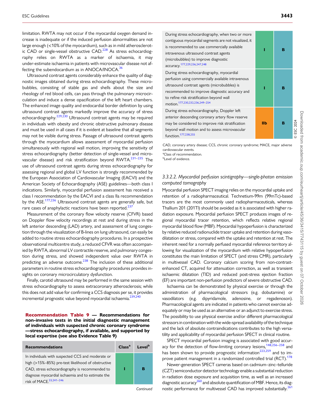
limitation. RWTA may not occur if the myocardial oxygen demand increase is inadequate or if the induced perfusion abnormalities are not large enough (<10% of the myocardium), such as in mild atherosclerotic CAD or single-vessel obstructive CAD.228 As stress echocardiography relies on RWTA as a marker of ischaemia, it may under-estimate ischaemia in patients with microvascular disease not affecting the subendocardium as in ANOCA/INOCA.36 Ultrasound contrast agents considerably enhance the quality of diagnostic images obtained during stress echocardiography. These microbubbles, consisting of stable gas and shells about the size and rheology of red blood cells, can pass through the pulmonary microcirculation and induce a dense opacification of the left heart chambers. The enhanced image quality and endocardial border definition by using ultrasound contrast agents markedly improve the accuracy of stress echocardiography.229,230 Ultrasound contrast agents may be required in individuals with obesity and chronic obstructive pulmonary disease and must be used in all cases if it is evident at baseline that all segments may not be visible during stress. Passage of ultrasound contrast agents through the myocardium allows assessment of myocardial perfusion simultaneously with regional wall motion, improving the sensitivity of stress echocardiography (better detection of single-vessel and microvascular disease) and risk stratification beyond RWTA.231–235 The use of ultrasound contrast agents during stress echocardiography for assessing regional and global LV function is strongly recommended by the European Association of Cardiovascular Imaging (EACVI) and the American Society of Echocardiography (ASE) guidelines—both class I indications. Similarly, myocardial perfusion assessment has received a class I recommendation by the EACVI and a class IIa recommendation by the ASE.177,236 Ultrasound contrast agents are generally safe, but rare cases of anaphylactic reactions have been reported.237 Measurement of the coronary flow velocity reserve (CFVR) based on Doppler flow velocity recordings at rest and during stress in the left anterior descending (LAD) artery, and assessment of lung congestion through the visualization of B-lines on lung ultrasound, can easily be added to routine stress echocardiography procedures. In a prospective observational multicentre study, a reduced CFVR was often accompanied by RWTA, abnormal LV contractile reserve, and pulmonary congestion during stress, and showed independent value over RWTA in predicting an adverse outcome.238 The inclusion of these additional parameters in routine stress echocardiography procedures provides insights on coronary microcirculatory dysfunction. Finally, carotid ultrasound may be performed in the same session with stress echocardiography to assess extracoronary atherosclerosis; while this does not add value for confirming a CCS diagnosis per se, it provides incremental prognostic value beyond myocardial ischaemia.239,240 3.3.2.2 Myocardial perfusion scintigraphy—single-photon emission

computed tomography Myocardial perfusion SPECT imaging relies on the myocardial uptake and retention of a radiopharmaceutical. Technetium-99m (99mTc)-based tracers are the most commonly used radiopharmaceuticals, whereas Thallium 201 (201Tl) should be avoided as it is associated with higher radiation exposure. Myocardial perfusion SPECT produces images of regional myocardial tracer retention, which reflects relative regional myocardial blood flow (MBF). Myocardial hypoperfusion is characterized by relative reduced radionuclide tracer uptake and retention during vasodilatation or stress, compared with the uptake and retention at rest. The inherent need for a normally perfused myocardial reference territory allowing for visualization of the myocardium with relative hypoperfusion constitutes the main limitation of SPECT (and stress CMR), particularly in multivessel CAD. Coronary calcium scoring from non-contrastenhanced CT, acquired for attenuation correction, as well as transient ischaemic dilatation (TID) and reduced post-stress ejection fraction (EF) are important non-perfusion predictors of severe obstructive CAD. Ischaemia can be demonstrated by physical exercise or through the administration of pharmacological stressors (e.g. dobutamine) or vasodilators (e.g. dipyridamole, adenosine, or regadenoson). Pharmacological agents are indicated in patients who cannot exercise adequately or may be used as an alternative or an adjunct to exercise stress. The possibility to use physical exercise and/or different pharmacological stressors in combination with the wide-spread availability of the technique and the lack of absolute contraindications contributes to the high versatility and applicability of myocardial perfusion SPECT in clinical routine. SPECT myocardial perfusion imaging is associated with good accuracy for the detection of flow-limiting coronary lesions,148,256–258 and has been shown to provide prognostic information223,259 and to improve patient management in a randomized controlled trial (RCT).178 Newer-generation SPECT cameras based on cadmium–zinc–telluride (CZT) semiconductor detector technology enable a substantial reduction in radiation dose exposure and acquisition time, as well as an increased diagnostic accuracy260 and absolute quantification of MBF. Hence, its diagnostic performance for multivessel CAD has improved substantially.261

Recommendation Table 9 — Recommendations for non-invasive tests in the initial diagnostic management of individuals with suspected chronic coronary syndrome —stress echocardiography, if available, and supported by local expertise (see also Evidence Table 9)

Recommendations Classa Levelb

In individuals with suspected CCS and moderate or high (>15%–85%) pre-test likelihood of obstructive CAD, stress echocardiography is recommended to diagnose myocardial ischaemia and to estimate the risk of MACE.33,241–246 I B

Continued During stress echocardiography, when two or more

contiguous myocardial segments are not visualized, it is recommended to use commercially available intravenous ultrasound contrast agents (microbubbles) to improve diagnostic accuracy.177,229,236,247,248 I B During stress echocardiography, myocardial perfusion using commercially available intravenous ultrasound contrast agents (microbubbles) is recommended to improve diagnostic accuracy and to refine risk stratification beyond wall motion.177,230,232,236,249–254 I B During stress echocardiography, Doppler left anterior descending coronary artery flow reserve may be considered to improve risk stratification beyond wall motion and to assess microvascular function.177,238,255 IIb B © ESC 2024

CAD, coronary artery disease; CCS, chronic coronary syndrome; MACE, major adverse cardiovascular events. aClass of recommendation. bLevel of evidence.

ESC Guidelines                                                                                                                                                                                          3443

Downloaded from academic.oup.com/eurheartj/article/45/36/3415/7743115 by guest on 07 October 2026