p.27 抽出テキスト
3.2.4 Chest X-ray

Chest X-ray is commonly utilized in the evaluation of patients experiencing chest pain. However, in the context of CCS, it does not yield specific information for accurate diagnosis or risk stratification. The test may provide assistance in assessing patients with suspected HF. Additionally, chest X-ray may prove beneficial in diagnosing pulmonary conditions that often co-exist with CAD, or in ruling out other potential causes of chest pain.

3.2.5 Ambulatory electrocardiogram monitoring

Ambulatory ECG monitoring can assist in evaluating patients with chest pain and palpitations. It can also help in detecting and evaluating silent myocardial ischaemia, as well as suspected VSA.192–194 3.3 STEP 3: Confirming the diagnosis

3.3.1. Anatomical imaging: coronary computed tomography angiography Through the intravenous (i.v.) injection of contrast agent, CCTA allows direct anatomical visualization of the coronary artery lumen and wall. CCTA offers a practical, non-invasive test, with proven diagnostic performance in detecting obstructive coronary artery stenoses when compared with ICA.32,148 Obstructive coronary stenoses have typically been defined using visual thresholds of either 50% or 70% diameter reduction. It is accepted that not all anatomical stenoses above such thresholds, especially those of moderate (50%–69%) stenosis severity, are haemodynamically or functionally significant195 or induce myocardial ischaemia.196 Depending on the clinical context, it may be necessary to complement CCTA with functional data either from non-invasive imaging techniques or from invasive angiography with fractional flow reserve (FFR) (see Section 3.3.3.2), when the haemodynamic consequence of a stenosis is deemed questionable for management options. While several earlier trials (publication date during or before 2016) reported a higher rate of downstream ICA in patients receiving CCTA compared with functional imaging,197 this was no longer observed in more recent trials (publication date after 2016). Moreover, increased downstream use of invasive procedures was linked to non-adherence to guideline recommendations as these procedures were used significantly less when the guidelines were adopted.198 Coronary computed tomography angiography-derived fractional flow reserve (FFR-CT) can complement CCTA by providing values of model-based computational FFR along the coronary tree. FFR-CT has shown good agreement with invasive FFR,199 and has clinical utility by reducing the number of unnecessary ICA procedures.200 However, in patients with severe disease at CCTA, FFR-CT has less impact on patient management.201 FFR-CT does not require pharmacological stress, additional contrast agent injection, or radiation exposure. FFR-CT, however, is not ubiquitous and depends on image quality. Nevertheless, the rejection rate is reported to be quite low in real-world data with newest-generation scanners.202–204

3.3.1.1 Computed tomography perfusion imaging

Computed tomography perfusion imaging, performed under pharmacological stress, has been validated against several reference standards, including single-photon computed tomography (SPECT) and

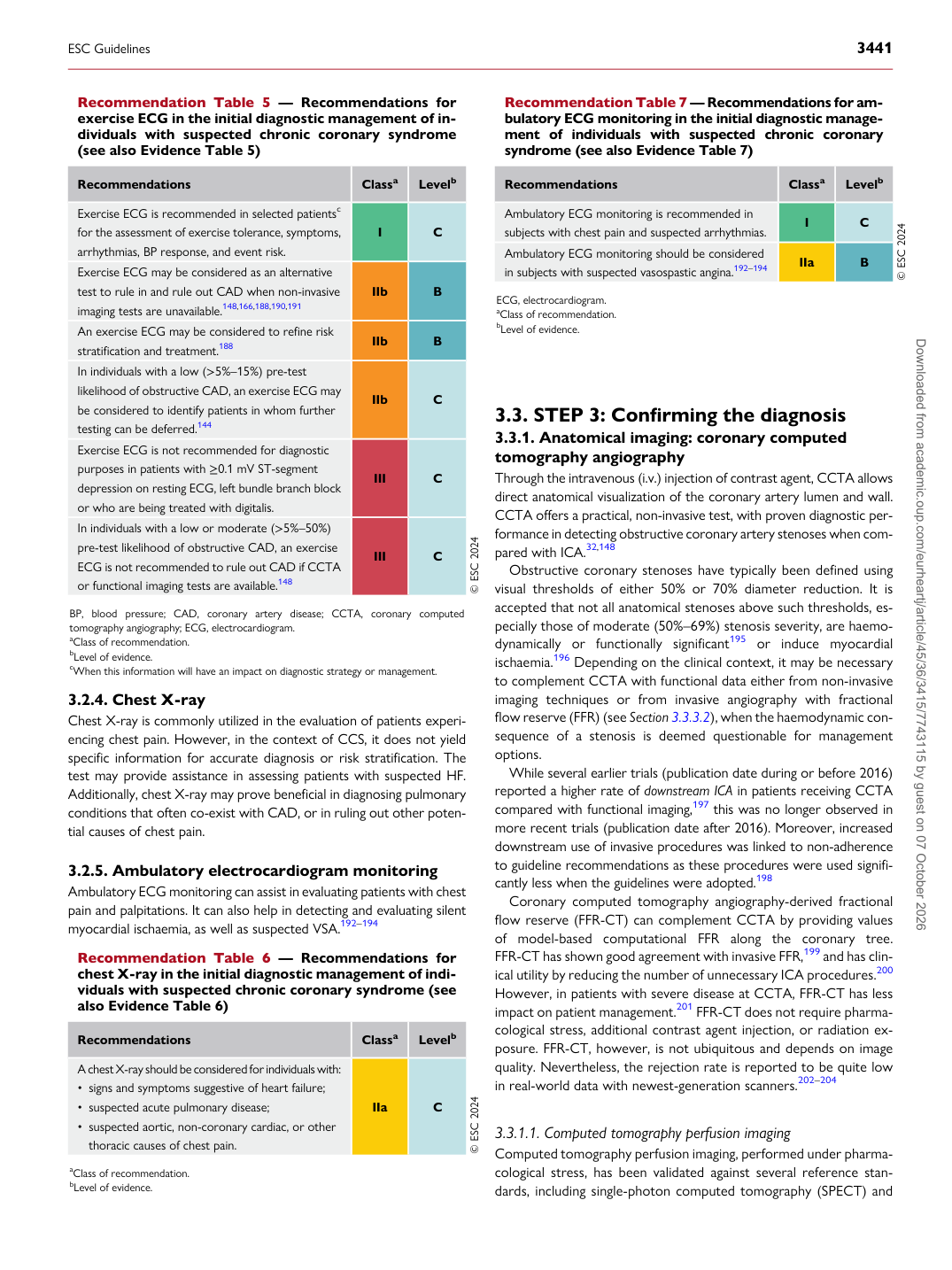
Recommendation Table 5 — Recommendations for exercise ECG in the initial diagnostic management of individuals with suspected chronic coronary syndrome (see also Evidence Table 5)

Recommendations Classa Levelb Exercise ECG is recommended in selected patientsc for the assessment of exercise tolerance, symptoms, arrhythmias, BP response, and event risk. I C Exercise ECG may be considered as an alternative test to rule in and rule out CAD when non-invasive imaging tests are unavailable.148,166,188,190,191 IIb B An exercise ECG may be considered to refine risk stratification and treatment.188 IIb B In individuals with a low (>5%–15%) pre-test likelihood of obstructive CAD, an exercise ECG may be considered to identify patients in whom further testing can be deferred.144 IIb C Exercise ECG is not recommended for diagnostic purposes in patients with ≥0.1 mV ST-segment depression on resting ECG, left bundle branch block or who are being treated with digitalis. III C In individuals with a low or moderate (>5%–50%) pre-test likelihood of obstructive CAD, an exercise ECG is not recommended to rule out CAD if CCTA or functional imaging tests are available.148 III C © ESC 2024

BP, blood pressure; CAD, coronary artery disease; CCTA, coronary computed tomography angiography; ECG, electrocardiogram. aClass of recommendation. bLevel of evidence. cWhen this information will have an impact on diagnostic strategy or management.

Recommendation Table 6 — Recommendations for chest X-ray in the initial diagnostic management of individuals with suspected chronic coronary syndrome (see also Evidence Table 6)

Recommendations Classa Levelb

A chest X-ray should be considered for individuals with: • signs and symptoms suggestive of heart failure; • suspected acute pulmonary disease; • suspected aortic, non-coronary cardiac, or other thoracic causes of chest pain. IIa C © ESC 2024 aClass of recommendation. bLevel of evidence. Recommendation Table 7 — Recommendations for ambulatory ECG monitoring in the initial diagnostic management of individuals with suspected chronic coronary syndrome (see also Evidence Table 7)

Recommendations Classa Levelb

Ambulatory ECG monitoring is recommended in subjects with chest pain and suspected arrhythmias. I C Ambulatory ECG monitoring should be considered in subjects with suspected vasospastic angina.192–194 IIa B © ESC 2024

ECG, electrocardiogram. aClass of recommendation. bLevel of evidence.

ESC Guidelines                                                                                                                                                                                          3441

Downloaded from academic.oup.com/eurheartj/article/45/36/3415/7743115 by guest on 07 October 2026