p.26 抽出テキスト
3.2.2 Transthoracic echocardiography and cardiac

magnetic resonance at rest An echocardiographic study will provide important information about cardiac function and anatomy. Patients with CCS have often preserved left ventricular ejection fraction (LVEF).167 A decreased LV function and/or regional wall motion abnormalities may increase the suspicion of ischaemic myocardial damage,167 and a pattern of LV dysfunction following the anatomical perfusion territory of the coronary arteries is typical in patients who have already had an MI.168,169 The detection of regional wall motion abnormalities can be challenging by visual assessment, and detection of early systolic lengthening, decreased systolic shortening, or post-systolic shortening by strain imaging techniques,170–172 or new parameters such as global myocardial work,173 may be helpful in individuals with apparently normal LV function but with clinical suspicion of CCS. Diastolic LV dysfunction has been reported to be an early sign of ischaemic myocardial dysfunction and may also be indicative of microvascular dysfunction.174,175 Echocardiography can help in detecting alternative causes of chest pain (e.g. pericarditis) and in diagnosing valvular heart diseases, ischaemic HF, and most cardiomyopathies,176 though these diseases may coexist with obstructive CAD. The use of an echocardiographic contrast agent can be helpful in patients with poor acoustic windows.177 Cardiac magnetic resonance (CMR) is an alternative in patients with suspected CAD when the echocardiogram (having used ultrasound contrast agent) is inconclusive.178 Cardiac magnetic resonance can assess global and regional function,179 and the use of late gadolinium enhancement (LGE) CMR can reveal a typical pattern of scarred myocardium in patients who have already experienced an MI.180 Moreover, CMR provides information on myocardial ischaemia through the evaluation of stress-induced perfusion defects.181 The strongest predictor of long-term survival is systolic LV function. Hence, risk stratification through the assessment of systolic LV function is useful in all symptomatic individuals with suspected CCS. Mortality increases as LVEF declines.182 Management of patients with either angina or HF symptoms, with reduced LVEF ≤40% or mildly reduced LVEF 41%–49%, is described in Section 4. 3.2.3 Exercise electrocardiogram testing

Exercise ECG testing is low cost, does not use ionizing radiation, is widely accessible, and remains an alternative for diagnostic testing depending on local resources and individual characteristics. The classical exercise ECG, involving graded exercise until the occurrence of fatigue, limiting chest pain or discomfort, significant ischaemic ECG changes, arrhythmias, excessive hypertension, a BP drop or after reaching 85% of the maximal predicted heart rate, has been the mainstay of the examination techniques used in clinical cardiology for assessing individuals with suspected CCS. Exercise ECG testing has a lower diagnostic performance of obstructive CAD compared with modern functional imaging and CCTA,148 which, therefore, should be preferred as a first-line test in subjects with suspected CCS. Several clinical trials have confirmed that a strategy based on anatomical34,146,187,188 or functional imaging189 simplifies the diagnosis, enables the targeting of preventive therapies and interventions, and potentially reduces the risk of MI compared with usual care based on exercise ECG. In addition, two randomized trials showed that patients reported fewer anginal complaints during follow-up when randomized to CCTA as an index investigation for stable chest pain compared with exercise ECG.145,146 Although the Scottish Computed Tomography of the Heart (SCOT-HEART) trial favoured CCTA as first-line test in CCS, a post hoc analysis suggested that abnormal results of exercise ECG remain a specific indicator of obstructive CAD, and are associated with future coronary revascularization and risk of MI.188 Exercise ECG testing with clearly abnormal results was most predictive for these outcomes; however, in a large proportion of individuals who underwent exercise ECG, particularly those with normal or inconclusive results, there was still a significant amount of unrecognized non-obstructive and obstructive CAD, which can be detected by additional CCTA imaging.188 In the WOMEN trial (What is the Optimal Method for Ischemia Evaluation of Women), including low-risk symptomatic women, exercise ECG was equally effective compared with exercise myocardial perfusion scintigraphy, with a similar 2-year incidence of major adverse cardiovascular events (MACE), defined as CAD death, or hospitalization for an ACS or HF, while providing significant diagnostic cost savings.190 Individuals exercising >10 metabolic equivalents with a negative exercise ECG and a low-risk Duke Treadmill Score have a good prognosis with limited need for downstream testing and revascularization.166,191 Patients with marked ischaemia at a low workload and a high-risk Duke Treadmill Score may benefit from further anatomical or functional testing. In regions with limited access to functional imaging or CCTA, or in individuals with a low (>5%–15%) pre-test likelihood of obstructive CAD,144 exercise ECG remains, therefore, useful for risk stratification and prognostication.144 Particularly, in subjects with a low (>5%– 15%) likelihood of obstructive CAD, a negative exercise ECG may help to down-classify patients into the very low likelihood (<5%) class, in whom further testing can be deferred.144 An exercise ECG is of no diagnostic value in patients with ECG abnormalities at rest that prevent interpretation of the ST-segment changes during stress (i.e. LBBB, paced rhythm, Wolff−Parkinson −White syndrome, ≥0.1 mV ST-segment depression on resting ECG, or treatment with digitalis). In patients with known CAD, exercise ECG may be considered in selected patients to complement their clinical evaluation for assessing symptoms, ST-segment changes, exercise tolerance, arrhythmias, BP response, and event risk. In summary, due to its low sensitivity (58%) and specificity (62%), exercise ECG testing has low diagnostic performance for the diagnosis of obstructive CAD148 and should mainly be used for risk stratification.

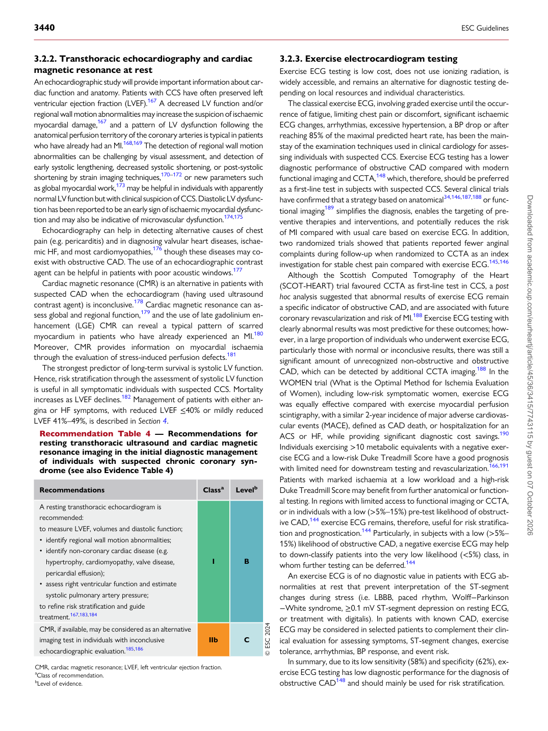
Recommendation Table 4 — Recommendations for resting transthoracic ultrasound and cardiac magnetic resonance imaging in the initial diagnostic management of individuals with suspected chronic coronary syndrome (see also Evidence Table 4)

Recommendations Classa Levelb

A resting transthoracic echocardiogram is recommended: to measure LVEF, volumes and diastolic function; • identify regional wall motion abnormalities; • identify non-coronary cardiac disease (e.g.

hypertrophy, cardiomyopathy, valve disease, pericardial effusion); • assess right ventricular function and estimate systolic pulmonary artery pressure; to refine risk stratification and guide treatment.167,183,184 I B CMR, if available, may be considered as an alternative imaging test in individuals with inconclusive echocardiographic evaluation.185,186 IIb C © ESC 2024

CMR, cardiac magnetic resonance; LVEF, left ventricular ejection fraction. aClass of recommendation. bLevel of evidence. Downloaded from academic.oup.com/eurheartj/article/45/36/3415/7743115 by guest on 07 October 2026