p.22 抽出テキスト
Recent-onset anginal symptoms with changing frequency or intensity should raise the suspicion that a coronary atherosclerotic plaque may be destabilizing. In these patients, the diagnostic algorithm recommended by the 2023 ESC Guidelines for the management of patients with acute coronary syndromes should be used to rule out an acute event.65 When investigating suspected CCS, it is important to perform a thorough physical examination that includes BP measurement and body mass index (BMI) calculation, to assess the presence of anaemia, hypertension, valvular heart disease, LV hypertrophy, or arrhythmias. It is also recommended to search for evidence of non-coronary vascular disease, which may be asymptomatic (palpation of peripheral pulses; auscultation of carotid and femoral arteries), and signs of other comorbid conditions, such as thyroid disease, renal disease, or diabetes. This should be used in the context of other clinical information, such as the presence of cough or stinging pain, making CCS less likely. One should also try to reproduce the symptoms by palpation and test the effect of sublingual nitroglycerine to classify the symptoms.

3.1.2 Basic testing: 12-lead electrocardiogram and

biochemistry Basic testing in individuals with suspected CCS includes a 12-lead ECG, standard laboratory tests, resting echocardiography, and, in selected patients, a chest X-ray, and a pulmonary function test if dyspnoea is the main symptom. Such tests can be done on an outpatient basis.

3.1.2.1 Electrocardiogram

The paradigm of diagnosing myocardial ischaemia has, for almost a century, been based on detecting repolarization abnormalities, mainly in the form of ST-segment depressions or T wave abnormalities. Thus, the resting 12-lead ECG remains an indispensable component of the initial evaluation of a patient with chest pain.67 A normal resting ECG is frequently recorded after an anginal attack. However, even in the absence of repolarization abnormalities, the ECG at rest may suggest CCS indirectly, through signs of previous MI (pathological Q or R waves) or conduction abnormalities [mainly left bundle branch block (LBBB) and impaired atrioventricular conduction]. Atrial fibrillation (AF) is not rarely associated with CCS.68 ST-segment depression during supraventricular tachyarrhythmias, however, is not a strong predictor of obstructive CAD.69–72

The ECG can be crucial for diagnosing transient myocardial ischaemia by recording dynamic ST-segment changes during ongoing angina. Vasospastic angina (VSA) should be suspected when observing typical transient ST-segment elevations or depressions with U-wave changes during an angina attack at rest.73 Long-term ambulatory ECG monitoring can be considered in selected patients to detect ischaemia during anginal episodes unrelated to physical activities. ECG changes suggesting ischaemia on ambulatory ECG monitoring are frequent in women but do not correlate with findings during stress testing.74 Ambulatory ECG monitoring may also reveal ‘silent’ ischaemia in patients with CCS, but therapeutic strategies targeting it have not demonstrated clear survival benefits.75,76

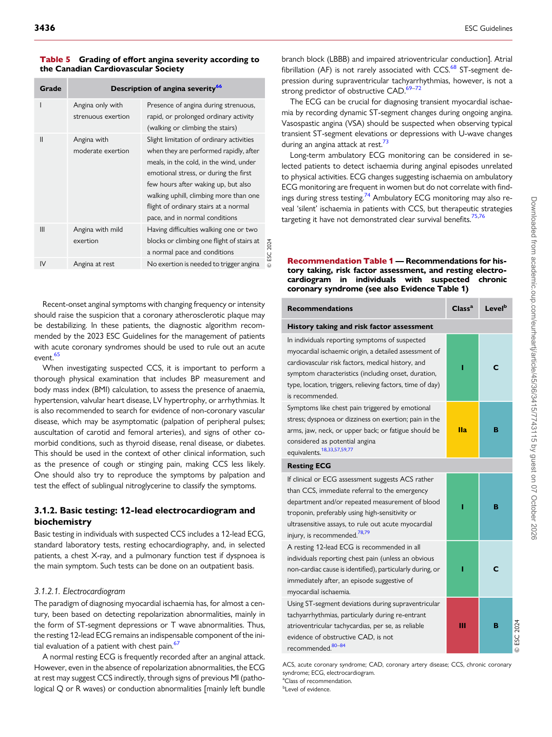
Table 5 Grading of effort angina severity according to the Canadian Cardiovascular Society

Grade Description of angina severity66

I Angina only with

strenuous exertion Presence of angina during strenuous,

rapid, or prolonged ordinary activity (walking or climbing the stairs)

II Angina with moderate exertion Slight limitation of ordinary activities when they are performed rapidly, after

meals, in the cold, in the wind, under emotional stress, or during the first few hours after waking up, but also walking uphill, climbing more than one flight of ordinary stairs at a normal pace, and in normal conditions

III Angina with mild exertion Having difficulties walking one or two blocks or climbing one flight of stairs at

a normal pace and conditions

IV Angina at rest No exertion is needed to trigger angina © ESC 2024 Recommendation Table 1 — Recommendations for history taking, risk factor assessment, and resting electrocardiogram in individuals with suspected chronic coronary syndrome (see also Evidence Table 1)

Recommendations Classa Levelb

History taking and risk factor assessment In individuals reporting symptoms of suspected myocardial ischaemic origin, a detailed assessment of cardiovascular risk factors, medical history, and symptom characteristics (including onset, duration, type, location, triggers, relieving factors, time of day) is recommended. I C Symptoms like chest pain triggered by emotional stress; dyspnoea or dizziness on exertion; pain in the arms, jaw, neck, or upper back; or fatigue should be considered as potential angina equivalents.18,33,57,59,77 IIa B Resting ECG If clinical or ECG assessment suggests ACS rather than CCS, immediate referral to the emergency department and/or repeated measurement of blood troponin, preferably using high-sensitivity or ultrasensitive assays, to rule out acute myocardial injury, is recommended.78,79 I B A resting 12-lead ECG is recommended in all individuals reporting chest pain (unless an obvious non-cardiac cause is identified), particularly during, or immediately after, an episode suggestive of myocardial ischaemia. I C Using ST-segment deviations during supraventricular tachyarrhythmias, particularly during re-entrant atrioventricular tachycardias, per se, as reliable evidence of obstructive CAD, is not recommended.80–84 III B © ESC 2024

ACS, acute coronary syndrome; CAD, coronary artery disease; CCS, chronic coronary syndrome; ECG, electrocardiogram. aClass of recommendation. bLevel of evidence. Downloaded from academic.oup.com/eurheartj/article/45/36/3415/7743115 by guest on 07 October 2026