p.60 抽出テキスト
Most consumer-based devices use photoplethysmography, and several large studies have been performed typically in low-risk individuals.633,1076,1117,1118 In an RCT of 5551 participants invited by their health insurer, smartphone-based photoplethysmography increased the odds of OAC-treated new AF by 2.12 (95% CI, 1.19–3.76; P = .01) compared with usual care.633 RCTs powered for assessment of clinical outcomes are still lacking for consumerbased AF screening. Further head-to-head comparisons between novel digital devices and those commonly used in healthcare settings are needed to establish their comparative effectiveness in the clinical setting and account for different populations and settings.1119 In a systematic review of smartphone-based photoplethysmography compared with a reference ECG, unrealistically high sensitivity and specificity were noted, likely due to small, lowquality studies with a high degree of patient selection bias.1120 Hence, when AF is suggested by a photoplethysmography device or any other screening tool, a single-lead or continuous ECG tracing of >30 s or 12-lead ECG showing AF analysed by a physician with expertise in ECG rhythm interpretation is recommended to establish a definitive diagnosis of AF.1091,1121–1125 The combination of big data and artificial intelligence (AI) is having an increasing impact on the field of electrophysiology. Algorithms have been created to improve automated AF diagnosis and several algorithms to aid diagnostics are being investigated.1046 However, the clinical performance and broad applicability of these solutions are not yet known. The use of AI may enable future treatment changes to be assessed with dynamic and continuous patient-directed monitoring using wearable devices.1126 There are still challenges in the field that need clarification, such as data acquisition, model performance, external validity, clinical implementation, algorithm interpretation, and confidence, as well as the ethical aspects.1127 10.3 Screening strategies for AF

Screening can be performed systematically, with an invitation issued to a patient, or opportunistically, at the time of an ad hoc meeting with a healthcare professional. Regardless of the mode of invitation, screening should be part of a structured programme1128 and is not the same as identification of AF during a routine healthcare visit or secondary to arrhythmia symptoms. Screening can be done at a single timepoint (snapshot of the heart rhythm), e.g. using pulse palpation or a 12-lead ECG. Screening can also be of an extended duration, i.e. prolonged, using either intermittent or continuous monitoring of heart rhythm. Most studies using an opportunistic strategy have screened for AF at a single timepoint with short duration (such as a single timepoint ECG), compared with systematic screening studies that have mainly used prolonged (repeated or continuous) rhythm assessment.1129 The optimal screening method will vary depending on the population being studied (Figure 16) (see Supplementary data online, Additional Evidence Table S32). More sensitive methods will detect more AF but may lead to an increased risk of false positives and an increased detection of low burden AF, whereas more specific methods result in less false positives, at the risk of missing AF. Invasive monitoring of heart rhythm in high-risk populations extended for several years has been shown to result in device-detected AF prevalence of around 30%, albeit most of whom have a low burden of AF.5,857,1130,1131 Pacemaker studies have shown that patients with a low burden of device-detected subclinical AF have a lower risk of ischaemic stroke.5,24,1131,1132 This has been confirmed in RCTs assessing DOAC use in patients with device-detected subclinical AF (see Section 6.1.1).5,281,282 The burden needed for device-detected subclinical AF to translate into stroke risk is not known, and further studies are clearly needed.1133,1134 Benefit and cost-effectiveness of screening are discussed in the Supplementary data online.

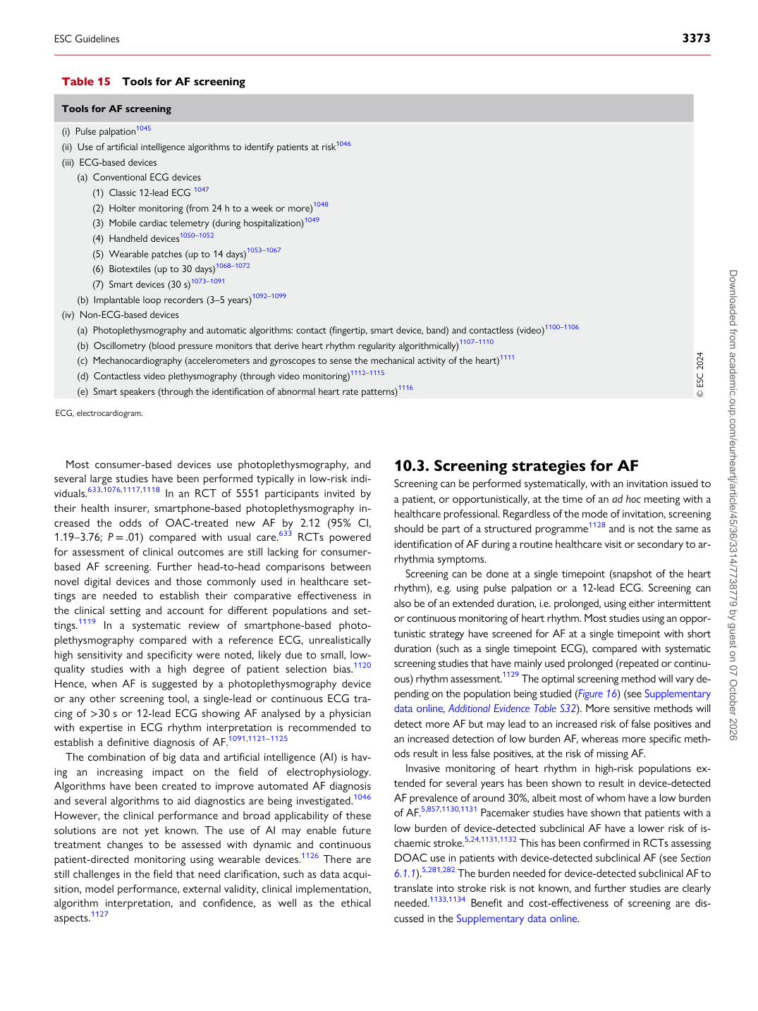
Table 15 Tools for AF screening

Tools for AF screening (i) Pulse palpation1045 (ii) Use of artificial intelligence algorithms to identify patients at risk1046 (iii) ECG-based devices

(a) Conventional ECG devices

(1) Classic 12-lead ECG 1047 (2) Holter monitoring (from 24 h to a week or more)1048 (3) Mobile cardiac telemetry (during hospitalization)1049 (4) Handheld devices1050–1052 (5) Wearable patches (up to 14 days)1053–1067 (6) Biotextiles (up to 30 days)1068–1072 (7) Smart devices (30 s)1073–1091 (b) Implantable loop recorders (3–5 years)1092–1099 (iv) Non-ECG-based devices

(a) Photoplethysmography and automatic algorithms: contact (fingertip, smart device, band) and contactless (video)1100–1106 (b) Oscillometry (blood pressure monitors that derive heart rhythm regularity algorithmically)1107–1110 (c) Mechanocardiography (accelerometers and gyroscopes to sense the mechanical activity of the heart)1111 (d) Contactless video plethysmography (through video monitoring)1112–1115 (e) Smart speakers (through the identification of abnormal heart rate patterns)1116 © ESC 2024

ECG, electrocardiogram.

ESC Guidelines                                                                                                                                                                                          3373

Downloaded from academic.oup.com/eurheartj/article/45/36/3314/7738779 by guest on 07 October 2026