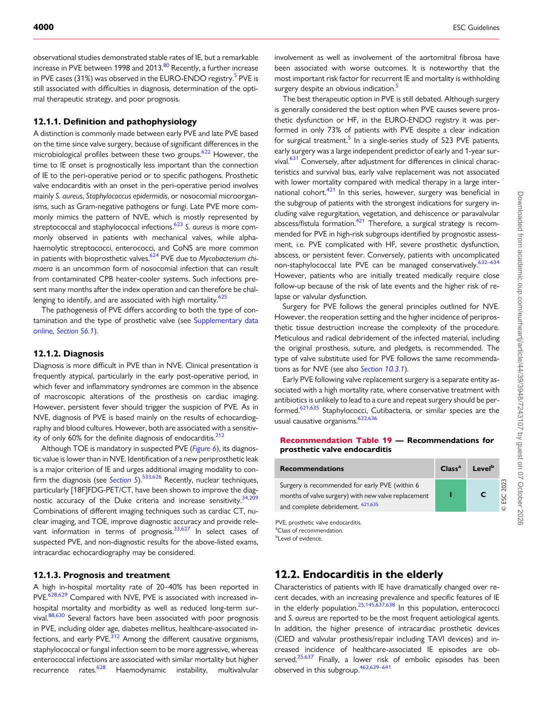
p.53 抽出テキスト
observational studies demonstrated stable rates of IE, but a remarkable increase in PVE between 1998 and 2013.80 Recently, a further increase in PVE cases (31%) was observed in the EURO-ENDO registry.5 PVE is still associated with difficulties in diagnosis, determination of the optimal therapeutic strategy, and poor prognosis.

12.1.1 Definition and pathophysiology

A distinction is commonly made between early PVE and late PVE based on the time since valve surgery, because of significant differences in the microbiological profiles between these two groups.622 However, the time to IE onset is prognostically less important than the connection of IE to the peri-operative period or to specific pathogens. Prosthetic valve endocarditis with an onset in the peri-operative period involves mainly S. aureus, Staphylococcus epidermidis, or nosocomial microorganisms, such as Gram-negative pathogens or fungi. Late PVE more commonly mimics the pattern of NVE, which is mostly represented by streptococcal and staphylococcal infections.623 S. aureus is more commonly observed in patients with mechanical valves, while alphahaemolytic streptococci, enterococci, and CoNS are more common in patients with bioprosthetic valves.624 PVE due to Mycobacterium chimaera is an uncommon form of nosocomial infection that can result from contaminated CPB heater-cooler systems. Such infections present many months after the index operation and can therefore be challenging to identify, and are associated with high mortality.625 The pathogenesis of PVE differs according to both the type of contamination and the type of prosthetic valve (see Supplementary data online, Section S6.1).

12.1.2 Diagnosis

Diagnosis is more difficult in PVE than in NVE. Clinical presentation is frequently atypical, particularly in the early post-operative period, in which fever and inflammatory syndromes are common in the absence of macroscopic alterations of the prosthesis on cardiac imaging. However, persistent fever should trigger the suspicion of PVE. As in NVE, diagnosis of PVE is based mainly on the results of echocardiography and blood cultures. However, both are associated with a sensitivity of only 60% for the definite diagnosis of endocarditis.212 Although TOE is mandatory in suspected PVE (Figure 6), its diagnostic value is lower than in NVE. Identification of a new periprosthetic leak is a major criterion of IE and urges additional imaging modality to confirm the diagnosis (see Section 5).533,626 Recently, nuclear techniques, particularly [18F]FDG-PET/CT, have been shown to improve the diagnostic accuracy of the Duke criteria and increase sensitivity.34,209 Combinations of different imaging techniques such as cardiac CT, nuclear imaging, and TOE, improve diagnostic accuracy and provide relevant information in terms of prognosis.33,627 In select cases of suspected PVE, and non-diagnostic results for the above-listed exams, intracardiac echocardiography may be considered.

12.1.3 Prognosis and treatment

A high in-hospital mortality rate of 20–40% has been reported in PVE.628,629 Compared with NVE, PVE is associated with increased inhospital mortality and morbidity as well as reduced long-term survival.88,630 Several factors have been associated with poor prognosis in PVE, including older age, diabetes mellitus, healthcare-associated infections, and early PVE.312 Among the different causative organisms, staphylococcal or fungal infection seem to be more aggressive, whereas enterococcal infections are associated with similar mortality but higher recurrence rates.628 Haemodynamic instability, multivalvular involvement as well as involvement of the aortomitral fibrosa have been associated with worse outcomes. It is noteworthy that the most important risk factor for recurrent IE and mortality is withholding surgery despite an obvious indication.5

The best therapeutic option in PVE is still debated. Although surgery is generally considered the best option when PVE causes severe prosthetic dysfunction or HF, in the EURO-ENDO registry it was performed in only 73% of patients with PVE despite a clear indication for surgical treatment.5 In a single-series study of 523 PVE patients, early surgery was a large independent predictor of early and 1-year survival.631 Conversely, after adjustment for differences in clinical characteristics and survival bias, early valve replacement was not associated with lower mortality compared with medical therapy in a large international cohort.421 In this series, however, surgery was beneficial in the subgroup of patients with the strongest indications for surgery including valve regurgitation, vegetation, and dehiscence or paravalvular abscess/fistula formation.421 Therefore, a surgical strategy is recommended for PVE in high-risk subgroups identified by prognostic assessment, i.e. PVE complicated with HF, severe prosthetic dysfunction, abscess, or persistent fever. Conversely, patients with uncomplicated non-staphylococcal late PVE can be managed conservatively.632–634 However, patients who are initially treated medically require close follow-up because of the risk of late events and the higher risk of relapse or valvular dysfunction. Surgery for PVE follows the general principles outlined for NVE. However, the reoperation setting and the higher incidence of periprosthetic tissue destruction increase the complexity of the procedure. Meticulous and radical debridement of the infected material, including the original prosthesis, suture, and pledgets, is recommended. The type of valve substitute used for PVE follows the same recommendations as for NVE (see also Section 10.3.1). Early PVE following valve replacement surgery is a separate entity associated with a high mortality rate, where conservative treatment with antibiotics is unlikely to lead to a cure and repeat surgery should be performed.621,635 Staphylococci, Cutibacteria, or similar species are the usual causative organisms.622,636

12.2 Endocarditis in the elderly

Characteristics of patients with IE have dramatically changed over recent decades, with an increasing prevalence and specific features of IE in the elderly population.25,145,637,638 In this population, enterococci and S. aureus are reported to be the most frequent aetiological agents. In addition, the higher presence of intracardiac prosthetic devices (CIED and valvular prosthesis/repair including TAVI devices) and increased incidence of healthcare-associated IE episodes are observed.25,637 Finally, a lower risk of embolic episodes has been observed in this subgroup.462,639–641

Recommendation Table 19 — Recommendations for prosthetic valve endocarditis

Recommendations Classa Levelb

Surgery is recommended for early PVE (within 6 months of valve surgery) with new valve replacement and complete debridement. 621,635 I C © ESC 2023

PVE, prosthetic valve endocarditis. aClass of recommendation. bLevel of evidence. Downloaded from academic.oup.com/eurheartj/article/44/39/3948/7243107 by guest on 07 October 2026