p.47 抽出テキスト
surgical spinal treatment should be considered.526 In patients with urgent indication for cardiac surgery, the presence of these lesions does not contraindicate the cardiac intervention. Spondylodiscitis does not appear to worsen the prognosis of patients with IE but delaying the diagnosis of IE in patients with spondylodiscitis is associated with poor prognosis.248,517–520

9.6.2 Rheumatological manifestations

The pathogenesis of rheumatological manifestations and musculoskeletal symptoms in IE is not well established. The probable immunologicalinflammatory aetiology of this clinical presentation is supported by a variety of antibodies and laboratory markers, the sterility of the synovial fluid, and the rapid resolution without sequelae.527 Myalgia and back pain are reported in 12–15% of cases. Arthralgia occurs in ∼10% of patients, sometimes sequentially affecting several joints. Slightly less often are symptoms of peripheral arthritis preferentially involving the major and proximal joints at the lower extremities.5,145,182,516 Sacroiliitis is less frequently observed (1% of cases) as well as polymyalgia rheumatic-like syndrome with pain and morning stiffness of the shoulders and hips, proximal muscle weakness (0.9% of cases), and cutaneous leucocytoclastic vasculitis (purpuric skin lesions, 3.6% of cases).182,527,528 Rheumatological manifestations and musculoskeletal symptoms show rapid and complete resolution with antibiotics and their presence does not impact on the prognosis of IE.182,529

9.7 Acute renal failure

Acute renal failure is a common complication of IE and is associated with increased morbidity and mortality as well as significant increase in length and cost of hospitalization.5,531–534 Additionally, renal failure is an independent predictor of poor outcome after cardiac surgery.417 However, acute renal failure should not be a reason to delay cardiac surgery. The EURO-ENDO registry reported that in patients with IE, acute renal failure was the second most common complication with an incidence of almost 18%.5 Some single-centre studies that specifically reported on the incidence of acute renal failure (using standardized criteria) in patients with IE reported that any degree of acute renal failure from mild to severe might be observed in 40–69% of cases.532,535,536

Severe renal failure requiring haemodialysis has been reported in 6% of patients with IE and it is associated with a very high risk of mortality (40%).537 Several factors may be responsible for the onset or worsening of renal dysfunction: (i) immune complex and vasculitic glomerulonephritis; (ii) renal infarction due to septic emboli;538,539 (iii) haemodynamic impairment in patients with HF; (iv) antibiotic and other drug toxicity (notably related to aminoglycosides, vancomycin, nafcillin, amoxicillin, oxacillin, concomitant use of non-steroidal anti-inflammatories, and/ or high dose loop diuretics); and (v) nephrotoxicity of contrast agents used for diagnostic imaging techniques.417,531,534,535,537,540 To reduce the incidence of acute renal failure, nephrotoxic antibiotics should be avoided if possible or, if not possible, serum levels (aminoglycosides and vancomycin) as well as creatinine should be closely monitored, and the optimal dose of medication should be periodically re-evaluated and discussed with the Endocarditis Team and a pharmacologist.536 Loop diuretics should also be used cautiously and other potentially nephrotoxic drugs, such as non-steroidal anti-inflammatories, should be avoided.536 Similarly, the use of nephrotoxic contrast agents for diagnostic imaging techniques should be carefully evaluated and avoided when possible. In patients with IE and a reduced glomerular filtration rate, contrast enhanced abdominal ultrasound or MRI are reasonable tests to diagnose embolization as cause of renal function impairment.541

10 Surgical therapy: principles and

methods

Surgery has been demonstrated to be an independent predictor of survival in many retrospective studies of IE patients under various clinical conditions and offers a potential curative therapy to select patient groups.5,250,403,404,421,436 Optimal management of such patients may lead to lower peri-operative complication rates and further potential benefits of surgical therapy.

10.1 Pre-operative and peri-operative

management 10.1.1. Coronary angiography When cardiac surgery becomes necessary in IE, assessment of coronary anatomy is recommended (see Recommendation Table 16). Classically, pre-operative coronary angiography is recommended for men >40 years, post-menopausal women, and in those with one or more cardiovascular risk factors or history of CAD.128 The presence of aortic valve vegetations may preclude invasive coronary angiography due to the risk of iatrogenic embolization.542,543 However, some studies have demonstrated the safety of performing invasive coronary angiography in the presence of aortic valve vegetations, particularly in patients without very large and mobile vegetations.193,544 Alternatively, coronary CTA can be used to rule out significant coronary obstructions. Furthermore, surgery may need to be conducted without detailed information on coronary anatomy in certain clinical conditions, particularly emergencies. Of note, a recent study questioned the need for coronary artery bypass grafting of non-critical lesions at the time of surgery for IE and suggested that such concomitant intervention may have a negative impact on peri-operative outcomes.545

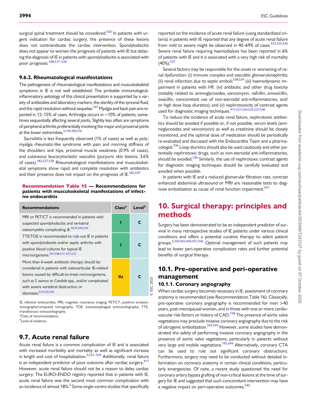
Recommendation Table 15 — Recommendations for patients with musculoskeletal manifestations of infective endocarditis

Recommendations Classa Levelb

MRI or PET/CT is recommended in patients with suspected spondylodiscitis and vertebral osteomyelitis complicating IE.30,32,206,524 I C TTE/TOE is recommended to rule out IE in patients with spondylodiscitis and/or septic arthritis with positive blood cultures for typical IE microorganisms.247,248,517–521,523 I C More than 6-week antibiotic therapy should be considered in patients with osteoarticular IE-related lesions caused by difficult-to-treat microorganisms, such as S. aureus or Candida spp., and/or complicated with severe vertebral destruction or abscesses.523,525,530 IIa C © ESC 2023

IE, infective endocarditis; MRI, magnetic resonance imaging; PET/CT, positron emission tomography/computed tomography, TOE, transoesophageal echocardiography; TTE, transthoracic echocardiography. aClass of recommendation. bLevel of evidence. Downloaded from academic.oup.com/eurheartj/article/44/39/3948/7243107 by guest on 07 October 2026