p.46 抽出テキスト
Nevertheless, one case series reported that it is safe to address the splenic abscess with splenectomy after valve repair.502 Alternatives to open splenectomy, i.e. percutaneous drainage507 and/or laparoscopic surgery,508 may be considered in patients with high surgical risk. After splenectomy, vaccination against encapsulated microorganisms (S. pneumoniae, N. meningitis, and Haemophilus spp.) is recommended.

9.4 Myocarditis and pericarditis

The actual prevalence of acute myocarditis in the setting of IE is unknown. Myocarditis will usually present in the form of acute HF and/ or ventricular arrhythmias indicating myocardial involvement in the inflammatory process most likely mediated by an immune mechanism. Differential diagnosis and exclusion of other potential complications are best assessed using echocardiography and cardiac MRI.509–511 Pericarditis is an infrequent complication of IE. In one retrospective series of 95 patients with aortic valve IE, 19% developed pericarditis usually related to ring abscess formation. The same authors also described a 12% rate of pericarditis associated with mitral valve IE.512,513 The pathophysiological mechanisms most commonly involved in IE-related pericarditis are the extension of inflammation from an infective aneurysm of the aortic root or valve ring abscess, an embolus in an extramural coronary artery, or the rupture of an infective aneurysm. In a recent large series of NVE, pericardial effusion was observed in 7.8% of patients and was associated with a higher risk of HF during admission. After adjusting for possible confounders, patients did not have a higher rate of surgery, and the presence of pericardial effusion was not associated with a higher in-hospital or 1-year mortality.513

9.5 Heart rhythm and conduction

disturbances Due to the critical anatomical relationship between heart valves and the conduction system, AVB may complicate the clinical presentation of IE. The atrioventricular node (AVN) and His bundle lie in close proximity to the insertion of the septal leaflet of the tricuspid valve, the aortic root (below the non-coronary and right coronary cusps), and the mitral annulus.514 A paravalvular abscess of these valves, especially of the aortic valve, may lead to AVB, and new electrocardiographic AVN conduction abnormalities are indicative of a paravalvular extension of the infection. In the EURO-ENDO registry, conduction abnormalities were observed at diagnosis in 11.5% of patients, including first-degree AVB in 8.1%, second-degree AVB in 0.6%, and third-degree AVB in 2.8% of cases.5 New-onset AVB caused by local extension of IE (i.e. abscess) is an indication for urgent cardiac surgery. Atrioventricular block may not only occur as a complication of paravalvular extension of the infection, but it may also develop as a consequence of valve surgery. In a series of 444 patients who survived cardiac surgery for IE,515 12.8% of patients required pacemaker implantation for AVB. Multivariable analysis identified that prolonged pre-operative PR and QRS intervals, S. aureus infection, presence of aortic root abscess, tricuspid valve involvement, and prior valvular surgery were independently associated with the need for post-operative pacemaker implantation. Pacemaker implantation should be considered in patients with surgery for valvular endocarditis and complete AVB if one or more of these risk factors is present.515 9.6 Musculoskeletal manifestations

9.6.1. Osteoarticular infective endocarditis-related infections Metastatic bone or joint IE-related lesions are relatively frequent due to the spread of the pathogen through the bloodstream and its subsequent tissue implantation. Although these lesions are considered an IE-related distal lesion or complication because infected valves are a continuous source of bacteraemia, it is often impossible to determine whether the primary infection is the valve or the osteoarticular infection. Overall, the incidence of osteoarticular infection among patients with IE is 6–8%, including bones, joints, and vertebral discs.5,145,247,516 The prevalence of spondylodiscitis ranges from 2% to 10% in patients with IE, including symptomatic and asymptomatic cases,248,517 while series of spontaneous spondylodiscitis have reported co-existing IE in up to 20–30% of patients.518–520 In general, the rate of IE is 10 times higher in patients with known spondylodiscitis. Therefore, in patients with a definite diagnosis of pyogenic spondylodiscitis and positive blood cultures, TTE/TOE is recommended to rule out IE.521 The most frequent microorganisms associated with spondylodiscitis are S. aureus, followed by Streptococcus spp., CoNS, and Enterococcus spp.247,248,305,517–523 The most common symptom of spondylodiscitis is back pain, although only 4% of patients with IE and back pain have spondylodiscitis.32,522 An MRI should be performed to accurately diagnose spondylodiscitis. Computed tomography can detect indirect signs of spondylodiscitis: loss of disc height, erosion/destruction of the endplates and vertebral bodies, and paravertebral soft tissue phlegmonous changes or abscess.206 Whole-body [18F]FDG-PET/CT can also identify spondylodiscitis.30,32,524 Indeed, spondylodiscitis is frequently detected as an incidental finding when PET/CT is performed for the diagnosis of PVE. Imaging techniques can also be helpful in guiding biopsies to obtain material for cultures in cases of suspected IE with negative blood cultures.206 Antibiotic treatment adapted to the antimicrobial susceptibility pattern is appropriate for most cases of spondylodiscitis. The outcome is usually favourable with the 4- to 6-week IE treatment course. Prolonged therapy is necessary in patients with IE caused by difficult-to-treat microorganisms, such as S. aureus or Candida spp., or in those with epidural or perivertebral abscesses.523,525 In patients with neurological deficits or severe spinal instability, the indication for

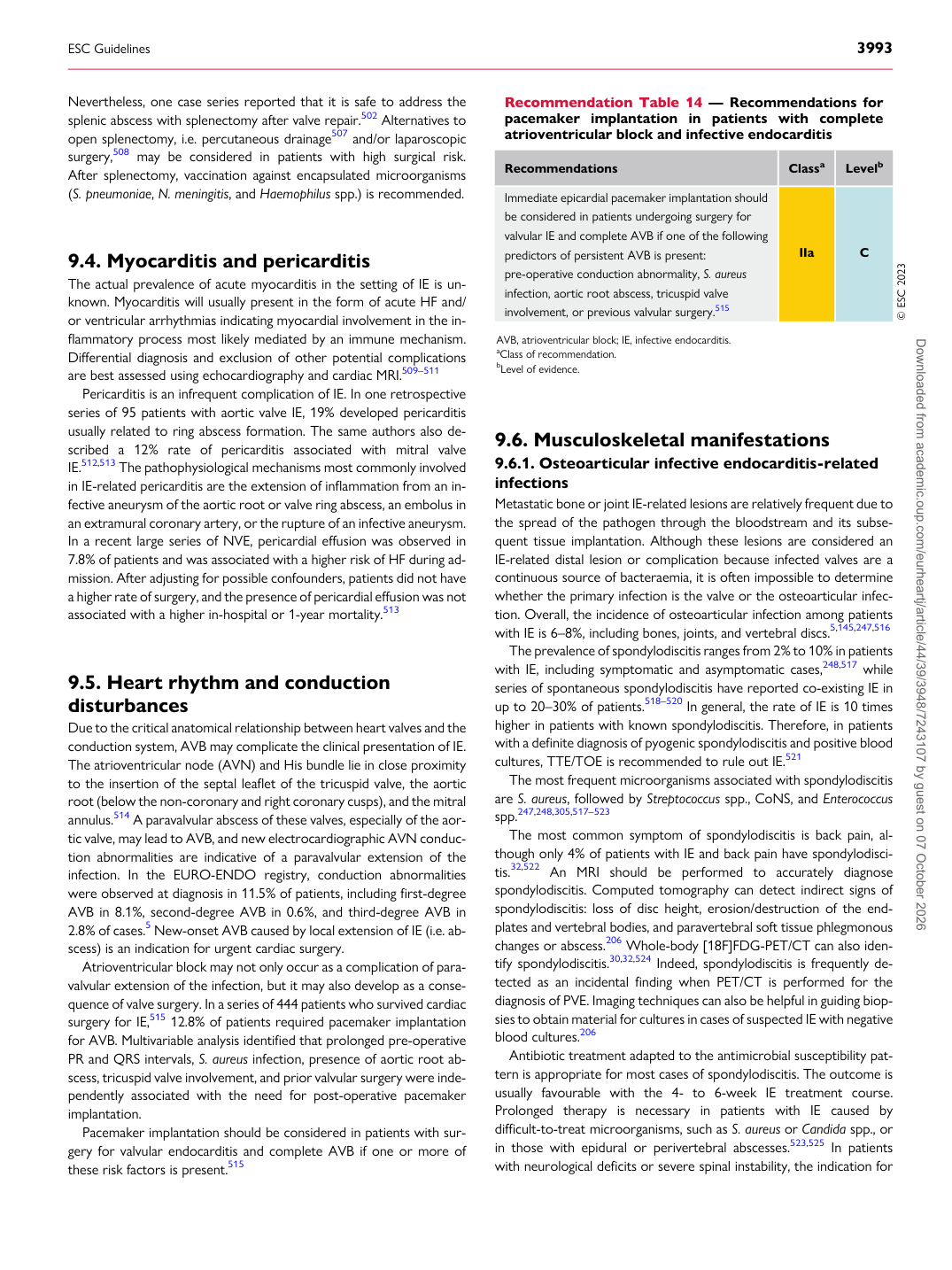
Recommendation Table 14 — Recommendations for pacemaker implantation in patients with complete atrioventricular block and infective endocarditis

Recommendations Classa Levelb

Immediate epicardial pacemaker implantation should be considered in patients undergoing surgery for valvular IE and complete AVB if one of the following predictors of persistent AVB is present: pre-operative conduction abnormality, S. aureus infection, aortic root abscess, tricuspid valve involvement, or previous valvular surgery.515 IIa C © ESC 2023

AVB, atrioventricular block; IE, infective endocarditis. aClass of recommendation. bLevel of evidence.

ESC Guidelines                                                                                                                                                                                          3993

Downloaded from academic.oup.com/eurheartj/article/44/39/3948/7243107 by guest on 07 October 2026