p.26 抽出テキスト
The reasons to justify the changes in the diagnostic criteria include:

5.5.1.1 Major criteria – microbiology

Enteroccus faecalis should be acknowledged as a typical endocarditis bacterium, regardless of the place of acquisition or the source of infection. Currently, the modified Duke criteria fail to identify 30% of E. faecalis definite IE. Using data from a prospective study of 344 patients with E. faecalis bacteraemia evaluated with echocardiography, Dahl et al. demonstrated that designating E. faecalis as a ‘typical’ endocarditis pathogen significantly improved the sensitivity to correctly identify definite IE, from 70% to 96%.245

5.5.1.2 Major criteria – imaging

(i) Diagnosis based on the presence of lesions characteristics of IE.

Anatomic lesions and increased [18F]FDG uptake or WBC accumulation can be depicted by nuclear imaging techniques and add

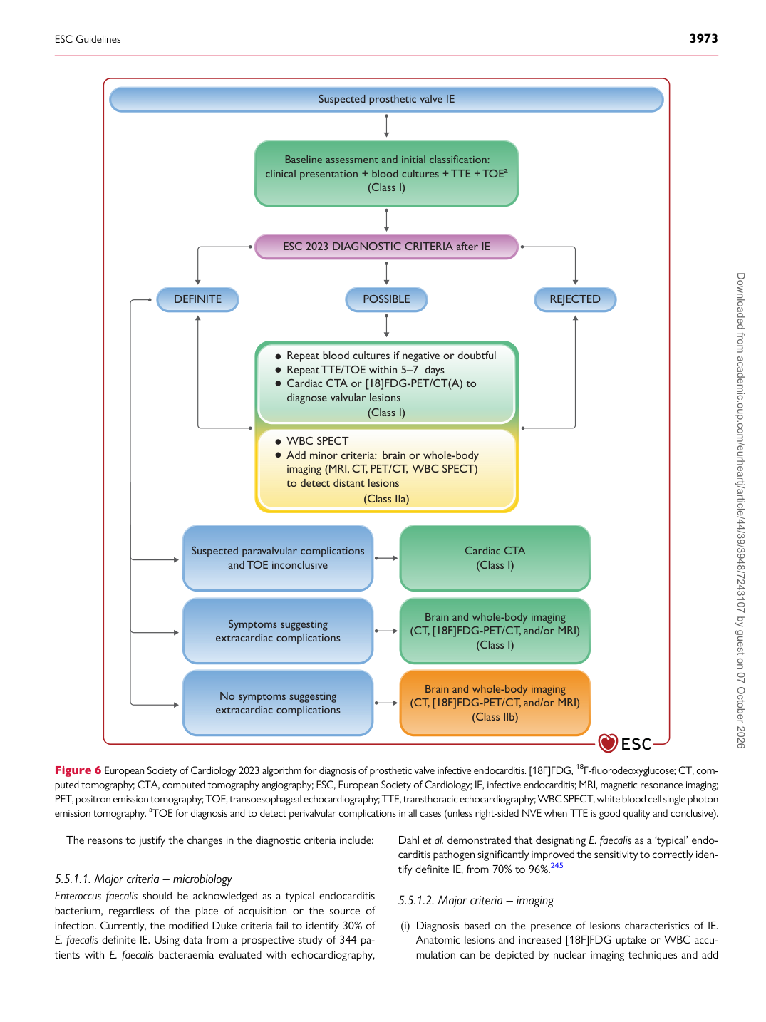
Repeat blood cultures if negative or doubtful Repeat TTE/TOE within 5–7  days Cardiac CTA or [18]FDG-PET/CT(A) to diagnose valvular lesions

WBC SPECT Add minor criteria:  brain or whole-body imaging (MRI, CT, PET/CT,  WBC SPECT) to detect distant lesions

Suspected paravalvular complications

and TOE inconclusive Cardiac CTA

(Class I)

No symptoms suggesting extracardiac complications Brain and whole-body imaging (CT, [18F]FDG-PET/CT, and/or MRI)

(Class IIb)

Symptoms suggesting extracardiac complications Brain and whole-body imaging (CT, [18F]FDG-PET/CT, and/or MRI)

(Class I)

Suspected prosthetic valve IE

ESC 2023 DIAGNOSTIC CRITERIA after IE

Baseline assessment and initial classification: clinical presentation + blood cultures + TTE + TOEa

(Class I)

POSSIBLE REJECTED DEFINITE

(Class I)

(Class IIa)

Figure 6 European Society of Cardiology 2023 algorithm for diagnosis of prosthetic valve infective endocarditis. [18F]FDG, 18F-fluorodeoxyglucose; CT, computed tomography; CTA, computed tomography angiography; ESC, European Society of Cardiology; IE, infective endocarditis; MRI, magnetic resonance imaging; PET, positron emission tomography; TOE, transoesophageal echocardiography; TTE, transthoracic echocardiography; WBC SPECT, white blood cell single photon emission tomography. aTOE for diagnosis and to detect perivalvular complications in all cases (unless right-sided NVE when TTE is good quality and conclusive).

ESC Guidelines                                                                                                                                                                                          3973

Downloaded from academic.oup.com/eurheartj/article/44/39/3948/7243107 by guest on 07 October 2026