p.25 抽出テキスト
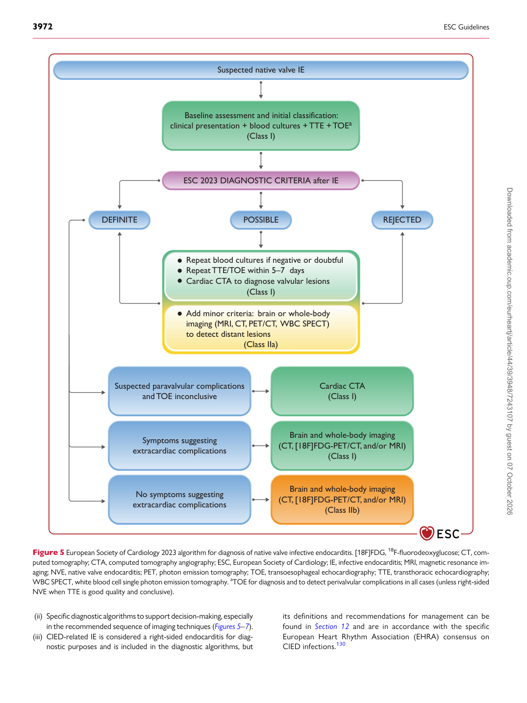
(ii) Specific diagnostic algorithms to support decision-making, especially in the recommended sequence of imaging techniques (Figures 5–7). (iii) CIED-related IE is considered a right-sided endocarditis for diag-

nostic purposes and is included in the diagnostic algorithms, but its definitions and recommendations for management can be found in Section 12 and are in accordance with the specific European Heart Rhythm Association (EHRA) consensus on CIED infections.130

Repeat blood cultures if negative or doubtful Repeat TTE/TOE within 5–7  days Cardiac CTA to diagnose valvular lesions

Add minor criteria:  brain or whole-body imaging (MRI, CT, PET/CT,  WBC SPECT) to detect distant lesions

Suspected paravalvular complications

and TOE inconclusive Cardiac CTA

(Class I)

No symptoms suggesting extracardiac complications Brain and whole-body imaging (CT, [18F]FDG-PET/CT, and/or MRI)

(Class IIb)

Symptoms suggesting extracardiac complications Brain and whole-body imaging (CT, [18F]FDG-PET/CT, and/or MRI)

(Class I)

Suspected native valve IE

ESC 2023 DIAGNOSTIC CRITERIA after IE

Baseline assessment and initial classification: clinical presentation + blood cultures + TTE + TOEa

(Class I)

POSSIBLE REJECTED DEFINITE

(Class I)

(Class IIa)

Figure 5 European Society of Cardiology 2023 algorithm for diagnosis of native valve infective endocarditis. [18F]FDG, 18F-fluorodeoxyglucose; CT, computed tomography; CTA, computed tomography angiography; ESC, European Society of Cardiology; IE, infective endocarditis; MRI, magnetic resonance imaging; NVE, native valve endocarditis; PET, photon emission tomography; TOE, transoesophageal echocardiography; TTE, transthoracic echocardiography; WBC SPECT, white blood cell single photon emission tomography. aTOE for diagnosis and to detect perivalvular complications in all cases (unless right-sided NVE when TTE is good quality and conclusive). Downloaded from academic.oup.com/eurheartj/article/44/39/3948/7243107 by guest on 07 October 2026