p.24 抽出テキスト
5.5 Diagnostic criteria

Since 2000, clinical, microbiological, and imaging findings have been integrated in the modified Duke criteria (see Supplementary data online, Table S6), which have demonstrated an overall sensitivity of 80% for IE.151 However, the clinical presentation of IE can be highly variable and some major limitations of the modified Duke criteria have become clear, particularly when prosthetic material is present (PVE, aortic grafts, cardiac devices, CHD). In these situations, echocardiography can be normal or inconclusive in up to 30% of cases despite the presence of IE.242–244 Therefore, the 2015 ESC diagnostic criteria introduced a multimodality imaging approach (echocardiography, cardiac/ whole-body CT, cerebral MRI, [18F]FDG-PET/CT, and WBC SPECT/ CT) to improve the diagnostic yield. This new approach has shown to be superior over the traditional diagnostic criteria.36–41,122,123,125,126,212

5.5.1 Modifications for the diagnosis of infective

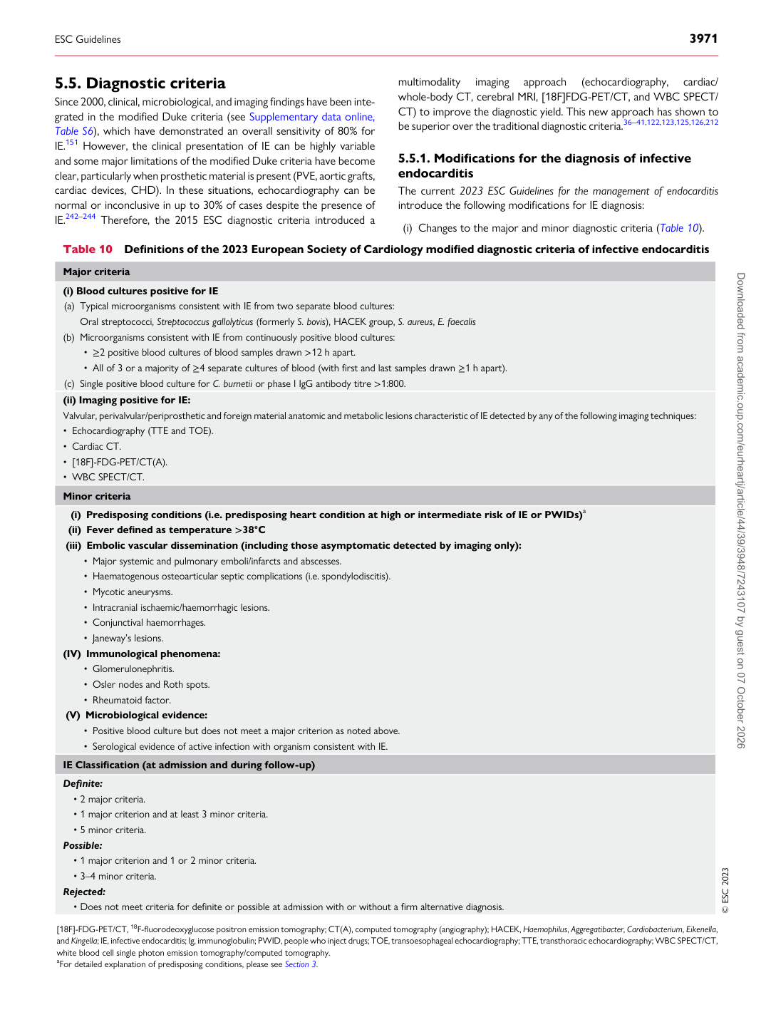
endocarditis The current 2023 ESC Guidelines for the management of endocarditis introduce the following modifications for IE diagnosis: (i) Changes to the major and minor diagnostic criteria (Table 10).

Table 10 Definitions of the 2023 European Society of Cardiology modified diagnostic criteria of infective endocarditis Major criteria (i) Blood cultures positive for IE (a) Typical microorganisms consistent with IE from two separate blood cultures:

Oral streptococci, Streptococcus gallolyticus (formerly S. bovis), HACEK group, S. aureus, E. faecalis (b) Microorganisms consistent with IE from continuously positive blood cultures:

• ≥2 positive blood cultures of blood samples drawn >12 h apart. • All of 3 or a majority of ≥4 separate cultures of blood (with first and last samples drawn ≥1 h apart). (c) Single positive blood culture for C. burnetii or phase I IgG antibody titre >1:800. (ii) Imaging positive for IE: Valvular, perivalvular/periprosthetic and foreign material anatomic and metabolic lesions characteristic of IE detected by any of the following imaging techniques: • Echocardiography (TTE and TOE). • Cardiac CT. • [18F]-FDG-PET/CT(A). • WBC SPECT/CT. Minor criteria

(i) Predisposing conditions (i.e. predisposing heart condition at high or intermediate risk of IE or PWIDs)a (ii) Fever defined as temperature >38°C (iii) Embolic vascular dissemination (including those asymptomatic detected by imaging only):

• Major systemic and pulmonary emboli/infarcts and abscesses. • Haematogenous osteoarticular septic complications (i.e. spondylodiscitis). • Mycotic aneurysms. • Intracranial ischaemic/haemorrhagic lesions. • Conjunctival haemorrhages. • Janeway’s lesions. (IV) Immunological phenomena:

• Glomerulonephritis. • Osler nodes and Roth spots. • Rheumatoid factor. (V) Microbiological evidence:

• Positive blood culture but does not meet a major criterion as noted above. • Serological evidence of active infection with organism consistent with IE. IE Classification (at admission and during follow-up) Definite:

• 2 major criteria. • 1 major criterion and at least 3 minor criteria. • 5 minor criteria. Possible:

• 1 major criterion and 1 or 2 minor criteria. • 3–4 minor criteria. Rejected:

• Does not meet criteria for definite or possible at admission with or without a firm alternative diagnosis. © ESC 2023 [18F]-FDG-PET/CT, 18F-fluorodeoxyglucose positron emission tomography; CT(A), computed tomography (angiography); HACEK, Haemophilus, Aggregatibacter, Cardiobacterium, Eikenella, and Kingella; IE, infective endocarditis; Ig, immunoglobulin; PWID, people who inject drugs; TOE, transoesophageal echocardiography; TTE, transthoracic echocardiography; WBC SPECT/CT, white blood cell single photon emission tomography/computed tomography. aFor detailed explanation of predisposing conditions, please see Section 3.

ESC Guidelines                                                                                                                                                                                          3971

Downloaded from academic.oup.com/eurheartj/article/44/39/3948/7243107 by guest on 07 October 2026