p.23 抽出テキスト
diagnosis of IE, as it can add a minor diagnostic criterion in patients without neurological symptoms with non-definitive IE diagnosis. Brain MRI can reclassify 25% of patients with an initially inconclusive diagnosis for IE to a more conclusive diagnosis, thereby leading to an earlier diagnosis.151 Cerebral microbleeds, found in 50–60% of patients with IE, are detected at gradient echo T2* sequences.200,202 Cerebral microbleeds should not be considered a minor criterion because there is no concordance with ischaemic lesions.203–205 (iii) Diagnosis of spine lesions. MRI is the diagnostic modality of choice

of spondylodiscitis and vertebral osteomyelitis with a diagnostic accuracy of 89–94%. MRI findings include vertebrae and disc oedema, paravertebral/epidural inflammation or abscess, bone erosion, and gadolinium enhancement of vertebrae and discs.32,206 It should be acknowledged that when MRI is performed too early, the rate of false-negative increases.207

5.4.4 Nuclear imaging positron emission

tomography/computed tomography (angiography) and single photon emission tomography/computed tomography Technical specifications of these imaging techniques are in the Supplementary data online, Section S2.2.2. The roles of nuclear imaging techniques in the diagnostic work-up of IE include: (i) Diagnosis of IE and cardiac complications. [18F]FDG-PET/CT and white

blood cell (WBC) single photon emission computed tomography (SPECT)/CT are recommended in suspected PVE in cases of inconclusive echocardiography. The most recent meta-analysis showed 86% sensitivity and 84% specificity for [18F]FDG-PET/CT in PVE.129 Additional evidence demonstrating the incremental diagnostic value of [18F]FDG-PET/CT and WBC SPECT/CT is summarized in the Supplementary data online, Section S2.2.2; Table S5.22,208–212

White blood cell SPECT/CT is an alternative nuclear imaging technique for the diagnosis of IE, when PET/CT is unavailable and inexperienced centres. The sensitivity of WBC SPECT/CT has been reported as 64–90% and the specificity as 36–100%; diagnostic ability significantly increases with the presence of periprosthetic abscesses.213–215 99mTechnetium-hexamethylpropyleneamine oxime (99mTc-HMPAO)-SPECT/CT helped to reduce the number of misdiagnosed IE cases classified in the ‘possible IE’ category by the modified Duke criteria by 27%.216 In cases of NVE, the sensitivity of PET/CT and SPECT/CT is low (about 31%) but with a higher specificity (around 98%).211 In NVE, the diagnosis of IE cannot be excluded in the absence of abnormal [18F]FDG uptake.217 The more frequent presence of valve vegetations in comparison with paravalvular involvement in NVE compared with PVE leads to reduced inflammatory response and subsequently lower [18F]FDG and WBC uptake. The lower sensitivity of [18F]FDG-PET/ CT is offset by other strengths of the technique, such as its ability to identify septic emboli when suspected.211,218–220 Electrocardiogram (ECG)-gated PET may further improve the diagnostic accuracy.221 Combining PET/CT acquisition with a CT angiography (PET/CTA) allows the detection of metabolic findings ([18F]FDG uptake distribution and intensity) and anatomical findings (IE-related lesions) within a single imaging procedure, resulting in the clinical clarification of indeterminate findings and change in the management of the patients.22,211 Such investigations may be particularly helpful in complex settings, such as patients with CHD222,223 and/or aortic grafts.22,224 (ii) Detection of distant lesions and sources of bacteraemia.

Whole-body [18F]FDG-PET/CT imaging is particularly useful in patients with a suspicion or proven IE to identify distant lesions, mycotic aneurysms, and the portal of entry of the infection.225,226 Septic emboli are typically located in the spleen, lungs (in rightsided IE), and kidneys, and metastatic infections in the intervertebral discs and/or the vertebral bone (spondylodiscitis) as well as in muscles and joints (septic arthritis) and liver.211,227,228 [18F] FDG-PET/CT is less suited to detect cerebral septic embolism and mycotic aneurysms of intracerebral arteries due to the high physiological uptake of [18F]FDG in the brain. (iii) Monitoring response to antimicrobial treatment with [18F]

FDG-PET/CT in patients with established IE and indication for surgery but who cannot be operated on due to unacceptable high risk and remain with long-term suppressive antibiotic treatment.137,184,229–236

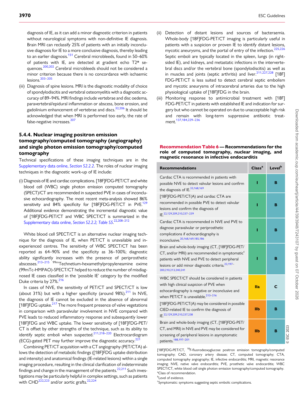
Recommendation Table 6 — Recommendations for the role of computed tomography, nuclear imaging, and magnetic resonance in infective endocarditis

Recommendations Classa Levelb

Cardiac CTA is recommended in patients with possible NVE to detect valvular lesions and confirm the diagnosis of IE.33,168,169 I B [18F]FDG-PET/CT(A) and cardiac CTA are recommended in possible PVE to detect valvular lesions and confirm the diagnosis of IE.22,129,209,210,237–239 I B Cardiac CTA is recommended in NVE and PVE to diagnose paravalvular or periprosthetic complications if echocardiography is inconclusive.20,168,169,185,186 I B Brain and whole-body imaging (CT, [18F]FDG-PET/ CT, and/or MRI) are recommended in symptomaticc patients with NVE and PVE to detect peripheral lesions or add minor diagnostic criteria.22,197– 200,210,213,240,241 I B

WBC SPECT/CT should be considered in patients with high clinical suspicion of PVE when echocardiography is negative or inconclusive and when PET/CT is unavailable.213–216 IIa C [18F]FDG-PET/CT(A) may be considered in possible CIED-related IE to confirm the diagnosis of IE.22,129,209,210,237,238 IIb B Brain and whole-body imaging (CT, [18F]FDG-PET/ CT, and MRI) in NVE and PVE may be considered for screening of peripheral lesions in asymptomatic patients.188,197–201 IIb B © ESC 2023 [18F]FDG-PET/CT, 18F-fluorodeoxyglucose positron emission tomography/computed tomography; CAD, coronary artery disease; CT, computed tomography; CTA, computed tomography angiography; IE, infective endocarditis; MRI, magnetic resonance imaging; NVE, native valve endocarditis; PVE, prosthetic valve endocarditis; WBC SPECT/CT, white blood cell single photon emission tomography/computed tomography. aClass of recommendation. bLevel of evidence. cSymptomatic: symptoms suggesting septic embolic complications. Downloaded from academic.oup.com/eurheartj/article/44/39/3948/7243107 by guest on 07 October 2026