p.22 抽出テキスト
5.4.2 Computed tomography

The indications for CT in patients with suspected or diagnosed IE include: (i) Diagnosis of IE and cardiac complications. Cardiac CT is more ac-

curate than TOE for diagnosing perivalvular and periprosthetic complications of IE (abscesses, pseudoaneurysms, and fistulae) and is recommended in both NVE and PVE if TOE is not conclusive or not feasible.33,168,169 In addition, cardiac CT can significantly influence subsequent surgical decision-making.20,185,186 Echocardiography continues to be superior for detecting valvular lesions, particularly small vegetations (<10 mm) which remain underdiagnosed by CT, but also leaflet perforations and fistulae (see Supplementary data online, Table S3).35,168,169 Cardiac CT should be acquired according to the recommendations of cardiac CT guidelines to ensure high diagnostic accuracy, and can be performed alone or in combination with PET.187 (ii) Detection of distant lesions and sources of bacteraemia. Whole-body and brain CT are useful for assessing IE systemic complications, including septic emboli. The detection of distant lesions adds a minor diagnostic criterion leading to a more conclusive diagnosis of definite or rejected IE, and can be relevant for decision-making.188 CT angiography can detect mycotic arterial aneurysms complicating IE in almost any site of the vascular tree,189,190 including the central nervous system (CNS). Although MRI is superior to CT for diagnosing neurological complications,191 CT may be more feasible in an emergency setting and is an acceptable alternative for the detection of neurological complications, with a sensitivity of 90% and specificity of 86% in the detection of ischaemic and haemorrhagic lesions.192 Finally, CT can also detect the extracardiac sources of the bacteraemia, including early neoplastic lesions, that may be important for patient management, and which need to be ideally addressed prior to undergoing heart valve surgery. However, CT does not replace the specific test indicated for the diagnosis of the extracardiac source of bacteraemia (i.e. colonoscopy in colon neoplasms). (iii) Pre-operative assessment. Cardiac CT is a valuable alternative for

non-invasive assessment of coronary artery disease (CAD) before cardiac surgery in patients with IE.193 (iv) Alternative diagnosis. In patients in whom IE is ruled out, or even in

doubtful patients with possible IE, an alternative diagnosis can be reached by whole-body CT, as it can help to detect alternative infectious foci. However, in these circumstances, an [18]FDG positron emission tomography/computed tomography (PET/CT) is the preferred imaging technique.194

5.4.3 Magnetic resonance imaging

The roles of MRI in the diagnostic work-up of IE include: (i) Diagnosis of IE and cardiac complications. The role of cardiac MRI to

diagnose IE is limited by the low spatial resolution (as compared with cardiac CT) and the signal void generated by some prostheses impairing the assessment of prosthetic valve anatomy and function.195,196 (ii) Diagnosis of neurological IE-related complications. MRI has higher

sensitivity than CT for the diagnosis of neurological lesions and, hence, increases the likelihood of detecting neurological complications in patients with IE. Patients with IE might present CNS lesions in up to 60–80% of cases,197 most of them corresponding to ischaemic lesions (50–80% of patients) that are often small and asymptomatic and do not impact on the decision-making.198 Other lesions that may influence the decision-making, such as parenchymal or subarachnoid haemorrhages, abscesses, or mycotic aneurysms, are found in <10% of patients.198–201 The systematic performance of brain MRI has shown to directly impact the

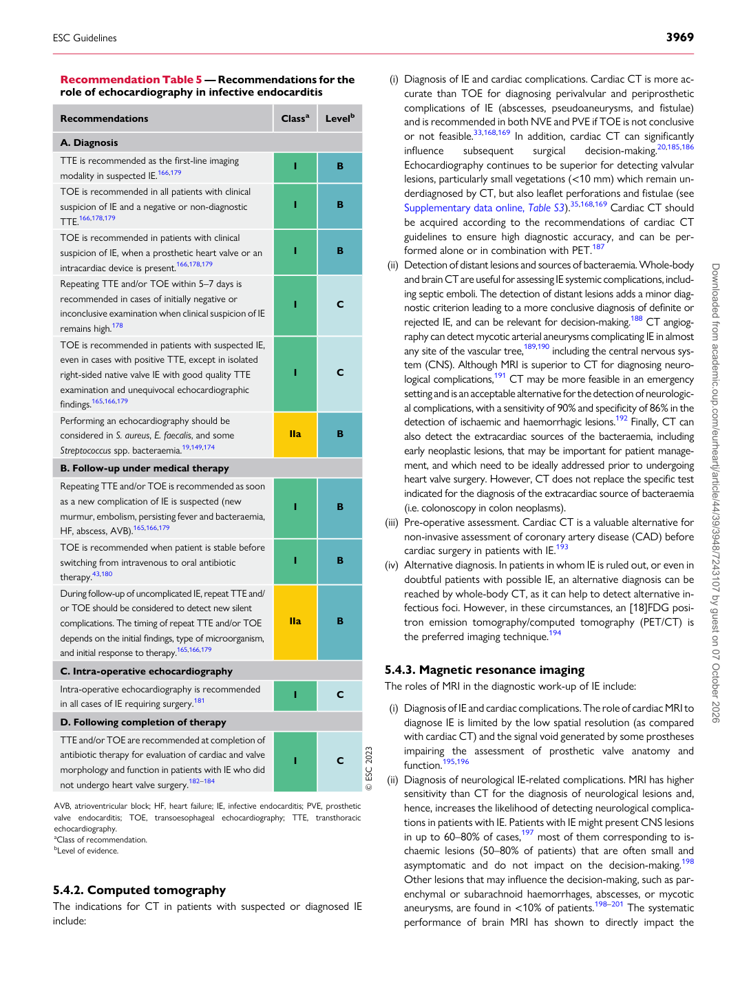
Recommendation Table 5 — Recommendations for the role of echocardiography in infective endocarditis

Recommendations Classa Levelb

A. Diagnosis TTE is recommended as the first-line imaging modality in suspected IE.166,179 I B TOE is recommended in all patients with clinical suspicion of IE and a negative or non-diagnostic TTE.166,178,179 I B TOE is recommended in patients with clinical suspicion of IE, when a prosthetic heart valve or an intracardiac device is present.166,178,179 I B Repeating TTE and/or TOE within 5–7 days is recommended in cases of initially negative or inconclusive examination when clinical suspicion of IE remains high.178 I C TOE is recommended in patients with suspected IE, even in cases with positive TTE, except in isolated right-sided native valve IE with good quality TTE examination and unequivocal echocardiographic findings.165,166,179 I C Performing an echocardiography should be considered in S. aureus, E. faecalis, and some Streptococcus spp. bacteraemia.19,149,174 IIa B B. Follow-up under medical therapy Repeating TTE and/or TOE is recommended as soon as a new complication of IE is suspected (new murmur, embolism, persisting fever and bacteraemia, HF, abscess, AVB).165,166,179 I B TOE is recommended when patient is stable before switching from intravenous to oral antibiotic therapy.43,180 I B During follow-up of uncomplicated IE, repeat TTE and/ or TOE should be considered to detect new silent complications. The timing of repeat TTE and/or TOE depends on the initial findings, type of microorganism, and initial response to therapy.165,166,179 IIa B C. Intra-operative echocardiography Intra-operative echocardiography is recommended in all cases of IE requiring surgery.181 I C D. Following completion of therapy TTE and/or TOE are recommended at completion of antibiotic therapy for evaluation of cardiac and valve morphology and function in patients with IE who did not undergo heart valve surgery.182–184 I C © ESC 2023

AVB, atrioventricular block; HF, heart failure; IE, infective endocarditis; PVE, prosthetic valve endocarditis; TOE, transoesophageal echocardiography; TTE, transthoracic echocardiography. aClass of recommendation. bLevel of evidence.

ESC Guidelines                                                                                                                                                                                          3969

Downloaded from academic.oup.com/eurheartj/article/44/39/3948/7243107 by guest on 07 October 2026