p.20 抽出テキスト
for antinuclear antibodies as well as antiphospholipid syndrome (APLs) (anticardiolipin antibodies [immunoglobulin (Ig)G] and anti-β2-glycoprotein 1 antibodies [IgG and IgM]) should be performed (although these antibodies may also be present in patients with proven IE).161,162 Pathological examination of resected tissue or embolic fragments remains the gold standard for IE diagnosis. All tissue samples that are excised during surgical valve debridement/resection must be collected in a sterile container without fixative or culture medium. Samples should be sent to the pathology department and the microbiology laboratory for the identification of microorganisms. On histological examination of excised valve tissue, patterns, and degrees of inflammation will vary depending on the infecting organism. Stains for bacteria, mycobacteria, and fungi may identify the microorganisms, and organism-specific immunohistochemical stains can be very useful for the final diagnosis. Importantly, histopathological analysis may facilitate the diagnosis of non-infectious causes of endocarditis, such as neoplastic and autoimmune causes.160

5.3.3 Proposed strategy for a microbiological

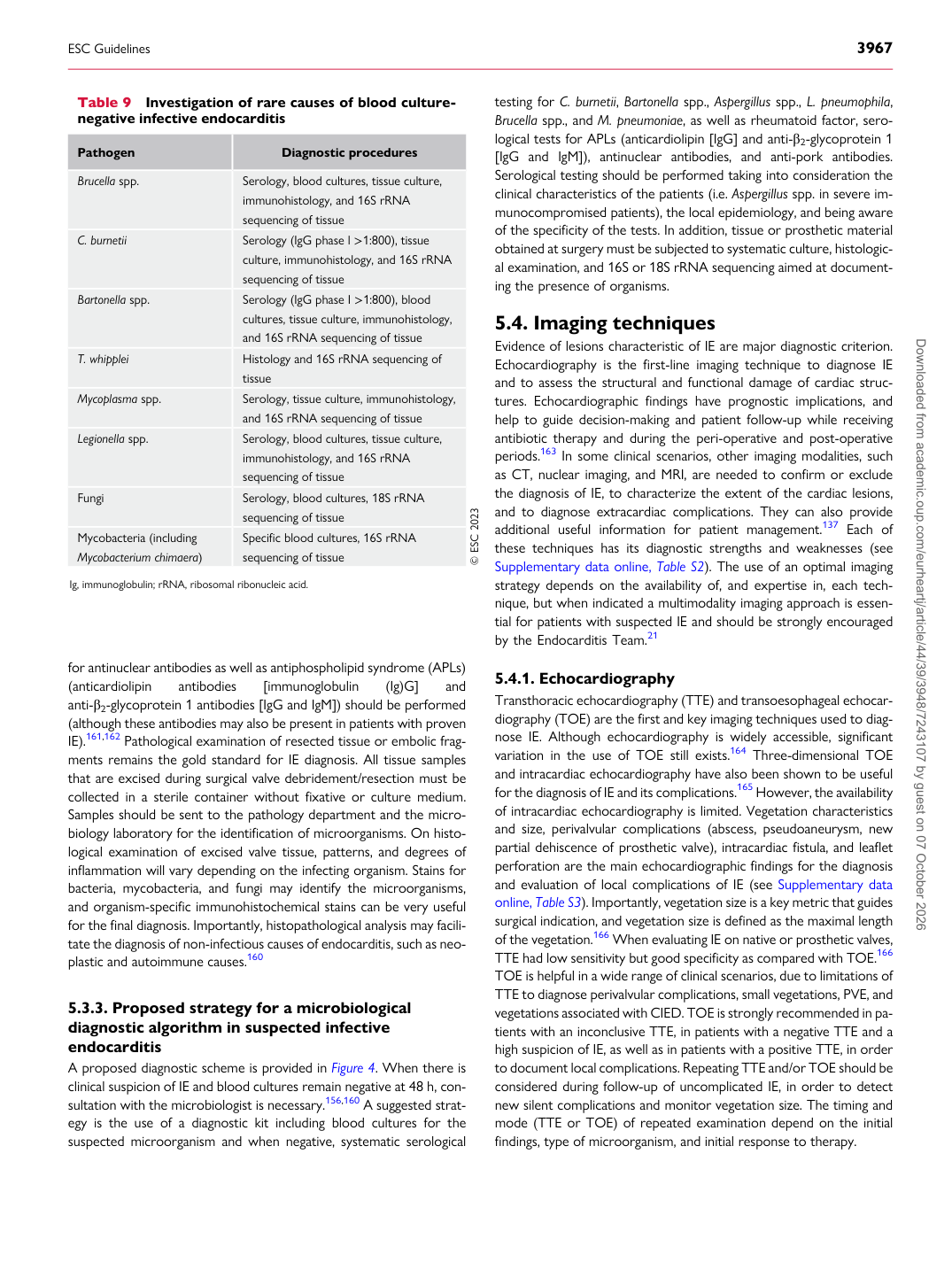
diagnostic algorithm in suspected infective endocarditis A proposed diagnostic scheme is provided in Figure 4. When there is clinical suspicion of IE and blood cultures remain negative at 48 h, consultation with the microbiologist is necessary.156,160 A suggested strategy is the use of a diagnostic kit including blood cultures for the suspected microorganism and when negative, systematic serological testing for C. burnetii, Bartonella spp., Aspergillus spp., L. pneumophila, Brucella spp., and M. pneumoniae, as well as rheumatoid factor, serological tests for APLs (anticardiolipin [IgG] and anti-β2-glycoprotein 1 [IgG and IgM]), antinuclear antibodies, and anti-pork antibodies. Serological testing should be performed taking into consideration the clinical characteristics of the patients (i.e. Aspergillus spp. in severe immunocompromised patients), the local epidemiology, and being aware of the specificity of the tests. In addition, tissue or prosthetic material obtained at surgery must be subjected to systematic culture, histological examination, and 16S or 18S rRNA sequencing aimed at documenting the presence of organisms.

5.4 Imaging techniques

Evidence of lesions characteristic of IE are major diagnostic criterion. Echocardiography is the first-line imaging technique to diagnose IE and to assess the structural and functional damage of cardiac structures. Echocardiographic findings have prognostic implications, and help to guide decision-making and patient follow-up while receiving antibiotic therapy and during the peri-operative and post-operative periods.163 In some clinical scenarios, other imaging modalities, such as CT, nuclear imaging, and MRI, are needed to confirm or exclude the diagnosis of IE, to characterize the extent of the cardiac lesions, and to diagnose extracardiac complications. They can also provide additional useful information for patient management.137 Each of these techniques has its diagnostic strengths and weaknesses (see Supplementary data online, Table S2). The use of an optimal imaging strategy depends on the availability of, and expertise in, each technique, but when indicated a multimodality imaging approach is essential for patients with suspected IE and should be strongly encouraged by the Endocarditis Team.21

5.4.1 Echocardiography

Transthoracic echocardiography (TTE) and transoesophageal echocardiography (TOE) are the first and key imaging techniques used to diagnose IE. Although echocardiography is widely accessible, significant variation in the use of TOE still exists.164 Three-dimensional TOE and intracardiac echocardiography have also been shown to be useful for the diagnosis of IE and its complications.165 However, the availability of intracardiac echocardiography is limited. Vegetation characteristics and size, perivalvular complications (abscess, pseudoaneurysm, new partial dehiscence of prosthetic valve), intracardiac fistula, and leaflet perforation are the main echocardiographic findings for the diagnosis and evaluation of local complications of IE (see Supplementary data online, Table S3). Importantly, vegetation size is a key metric that guides surgical indication, and vegetation size is defined as the maximal length of the vegetation.166 When evaluating IE on native or prosthetic valves, TTE had low sensitivity but good specificity as compared with TOE.166 TOE is helpful in a wide range of clinical scenarios, due to limitations of TTE to diagnose perivalvular complications, small vegetations, PVE, and vegetations associated with CIED. TOE is strongly recommended in patients with an inconclusive TTE, in patients with a negative TTE and a high suspicion of IE, as well as in patients with a positive TTE, in order to document local complications. Repeating TTE and/or TOE should be considered during follow-up of uncomplicated IE, in order to detect new silent complications and monitor vegetation size. The timing and mode (TTE or TOE) of repeated examination depend on the initial findings, type of microorganism, and initial response to therapy.

Table 9 Investigation of rare causes of blood culturenegative infective endocarditis

Pathogen Diagnostic procedures

Brucella spp. Serology, blood cultures, tissue culture,

immunohistology, and 16S rRNA sequencing of tissue

C. burnetii Serology (IgG phase l >1:800), tissue culture, immunohistology, and 16S rRNA

sequencing of tissue

Bartonella spp. Serology (IgG phase I >1:800), blood

cultures, tissue culture, immunohistology, and 16S rRNA sequencing of tissue

T. whipplei Histology and 16S rRNA sequencing of

tissue

Mycoplasma spp. Serology, tissue culture, immunohistology,

and 16S rRNA sequencing of tissue

Legionella spp. Serology, blood cultures, tissue culture,

immunohistology, and 16S rRNA sequencing of tissue

Fungi Serology, blood cultures, 18S rRNA sequencing of tissue Mycobacteria (including Mycobacterium chimaera) Specific blood cultures, 16S rRNA sequencing of tissue © ESC 2023

Ig, immunoglobulin; rRNA, ribosomal ribonucleic acid.

ESC Guidelines                                                                                                                                                                                          3967

Downloaded from academic.oup.com/eurheartj/article/44/39/3948/7243107 by guest on 07 October 2026