p.18 抽出テキスト
multimodality imaging are key in the Endocarditis Team. In addition, radiology and nuclear medicine specialists with expertise on clinical cardiovascular imaging should be available whenever indicated.22,31,129 The Endocarditis Team must meet on a frequent basis and work with standard operating procedures and the clinical governance arrangements defined locally.128,136 Although the decision of timing is left to the discretion of the local team, a weekly meeting is to be considered. In Referring Centres, i.e. those without a cardiovascular surgical team, the treating physician diagnosing IE should consult with a specialist in infectious diseases (or an internal medicine specialist with expertise in infectious diseases) and the microbiologist.136 In addition, a cardiologist with expertise in valvular heart disease and cardiac imaging should be present to provide the initial and subsequent evaluations with echocardiography. Information of the strains of the isolated microorganisms, usually kept for 7–15 days, should be provided to the Heart Valve Centre if requested. Communication between Referring Centres and the Heart Valve Centres should be facilitated with digital solutions that enable reliable data sharing. Early referral to the Heart Valve Centre for further diagnostic testing and clinical management should be available when deemed necessary (Figure 3). When there is evidence of failure to respond to the antibiotic therapy or there are complications related to valvular tissue destruction, the Referring Centre should consult the Heart Valve Centre. The Endocarditis Team of the Heart Valve Centre should share protocols with the physicians from the referring hospitals and should facilitate their continuing education.136 A critical aspect of the Endocarditis Team decision-making process is defining when a patient must be transferred to a Heart Valve Centre to expedite advanced diagnostics and therapy. The indications for transfer are comprehensive, to facilitate interhospital communication and avoid delaying therapy to improve prognosis.

5 Diagnosis

The diagnosis of IE is based on a clinical suspicion supported by consistent microbiological data and the documentation of IE-related cardiac lesions by imaging techniques. Evidence of involvement of cardiac valves (native or prosthetic) or prosthetic intracardiac material is a major diagnostic criterion of IE. Echocardiography is the first-line diagnostic imaging technique. Other imaging modalities such as CT, nuclear imaging, and magnetic resonance imaging (MRI) are currently part of the diagnostic strategy of suspected IE, given their ability to provide key information to confirm IE diagnosis, to assess local IE complications as well as IE-related distant lesions, and to identify the original source of bacteraemia in patients who develop secondary IE.137 Beyond diagnosis of IE, imaging findings also have prognostic implications.

5.1 Clinical features

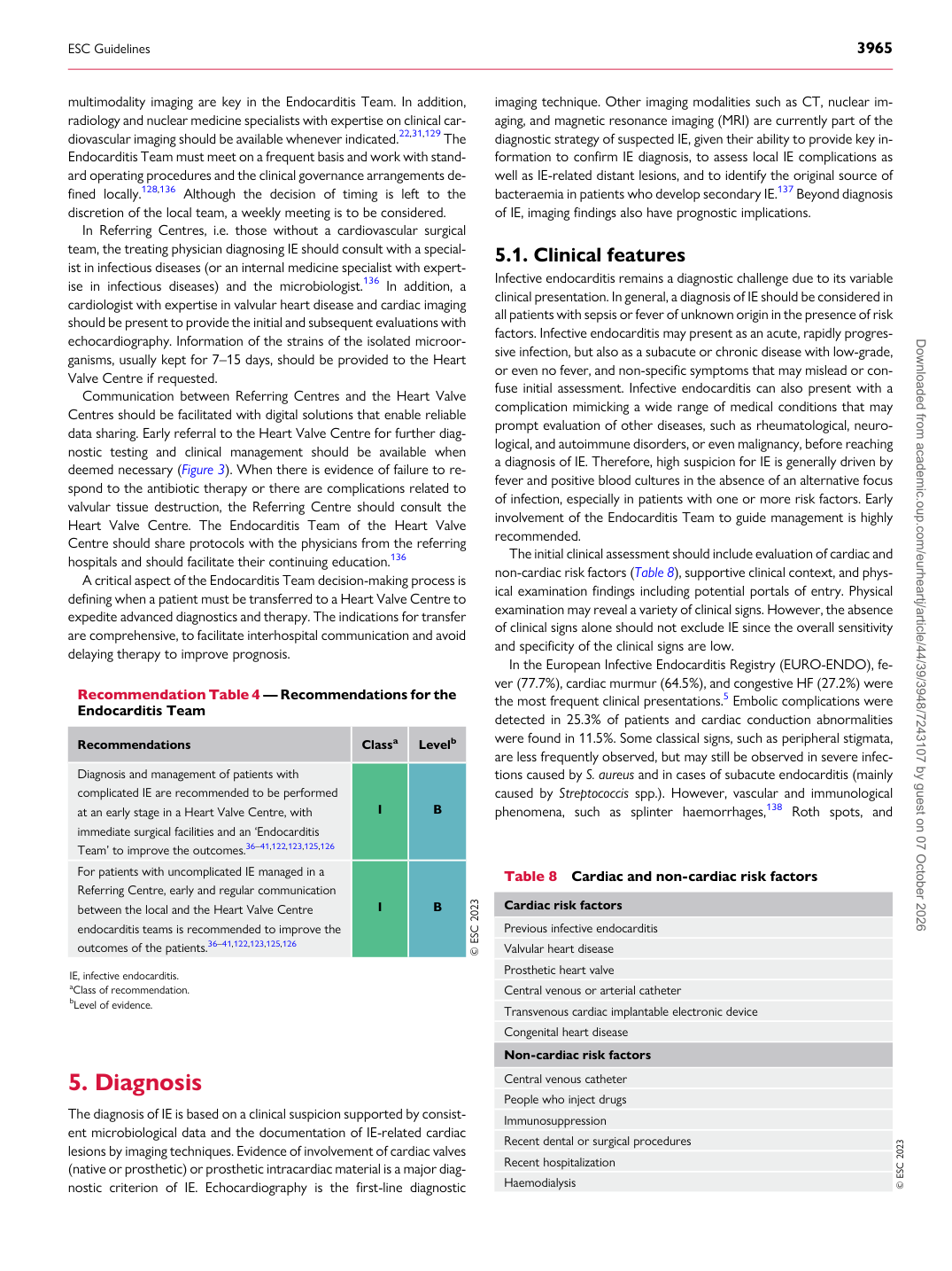
Infective endocarditis remains a diagnostic challenge due to its variable clinical presentation. In general, a diagnosis of IE should be considered in all patients with sepsis or fever of unknown origin in the presence of risk factors. Infective endocarditis may present as an acute, rapidly progressive infection, but also as a subacute or chronic disease with low-grade, or even no fever, and non-specific symptoms that may mislead or confuse initial assessment. Infective endocarditis can also present with a complication mimicking a wide range of medical conditions that may prompt evaluation of other diseases, such as rheumatological, neurological, and autoimmune disorders, or even malignancy, before reaching a diagnosis of IE. Therefore, high suspicion for IE is generally driven by fever and positive blood cultures in the absence of an alternative focus of infection, especially in patients with one or more risk factors. Early involvement of the Endocarditis Team to guide management is highly recommended. The initial clinical assessment should include evaluation of cardiac and non-cardiac risk factors (Table 8), supportive clinical context, and physical examination findings including potential portals of entry. Physical examination may reveal a variety of clinical signs. However, the absence of clinical signs alone should not exclude IE since the overall sensitivity and specificity of the clinical signs are low. In the European Infective Endocarditis Registry (EURO-ENDO), fever (77.7%), cardiac murmur (64.5%), and congestive HF (27.2%) were the most frequent clinical presentations.5 Embolic complications were detected in 25.3% of patients and cardiac conduction abnormalities were found in 11.5%. Some classical signs, such as peripheral stigmata, are less frequently observed, but may still be observed in severe infections caused by S. aureus and in cases of subacute endocarditis (mainly caused by Streptococcis spp.). However, vascular and immunological phenomena, such as splinter haemorrhages,138 Roth spots, and

Recommendation Table 4 — Recommendations for the Endocarditis Team

Recommendations Classa Levelb

Diagnosis and management of patients with complicated IE are recommended to be performed at an early stage in a Heart Valve Centre, with immediate surgical facilities and an ‘Endocarditis Team’ to improve the outcomes.36–41,122,123,125,126 I B For patients with uncomplicated IE managed in a Referring Centre, early and regular communication between the local and the Heart Valve Centre endocarditis teams is recommended to improve the outcomes of the patients.36–41,122,123,125,126 I B © ESC 2023

IE, infective endocarditis. aClass of recommendation. bLevel of evidence. Table 8 Cardiac and non-cardiac risk factors

Cardiac risk factors Previous infective endocarditis Valvular heart disease Prosthetic heart valve Central venous or arterial catheter Transvenous cardiac implantable electronic device Congenital heart disease Non-cardiac risk factors Central venous catheter People who inject drugs Immunosuppression Recent dental or surgical procedures Recent hospitalization Haemodialysis © ESC 2023

ESC Guidelines                                                                                                                                                                                          3965

Downloaded from academic.oup.com/eurheartj/article/44/39/3948/7243107 by guest on 07 October 2026