p.12 抽出テキスト
3 Prevention

3.1 Rationale

The development of IE usually requires several conditions, including the presence of predisposing risk factors (i.e. a surface/structure that could be colonized by bacteria), pathogens entering the bloodstream, and the competence of the host’s immune response. The role of predisposing risk factors has been recently underscored by Thornhill et al.47 Predisposing risk factors conveying a moderate and high risk of IE had an incidence of 280 and 497 cases per 100 000 subjects per year, respectively.47 The portals of entry of bacteria/fungi are variable and include: (i) infections of the skin, oral cavity, gastrointestinal, or genitourinary system; (ii) direct inoculation in people who inject drugs (PWID), or by any unsafe or unprotected vascular puncture; (iii) healthcare exposure (including a variety of invasive diagnostic or therapeutic procedures, such as transcatheter or surgical techniques).6,11,48–50 The oral cavity is colonized by relevant commensal flora, including oral group streptococci, and represents an important entry port. Oral surgery procedures (including all extractions, periodontal surgery, implant surgery, and oral biopsies) and dental procedures that involve manipulation of the gingival or periapical region of the teeth are considered at high risk of causing bacteraemia.11,48,49,51

Successful antibiotic prophylaxis assumes that reducing the bacteraemia associated with medical procedures will lead to a reduced risk of IE. This concept was supported by a few animal models and observational studies that led to the recommendation for antibiotic prophylaxis in a large number of patients with predisposing cardiac conditions undergoing a wide range of procedures.4,14,52–60 However, systematic use of antibiotic prophylaxis has been questioned based on several considerations, the most important being the lack of randomized clinical trials (RCTs) demonstrating the efficacy of antibiotic prophylaxis prior to medical procedures in preventing IE. Such trials would entail enrolment of a very large number of individuals and prolonged follow-up, making the feasibility of such studies improbable. Furthermore, since the standard of care for high-risk individuals is antibiotic prophylaxis (to date, mostly before invasive oro-dental procedures), there may not be sufficient equipoise to perform such RCTs. Finally, the costs of performing such trials have been considered unacceptable.61 To overcome these limitations, population-based studies

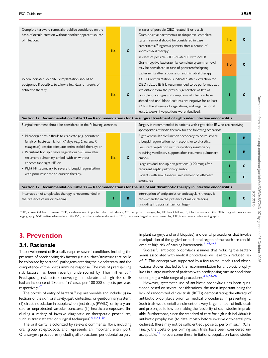
Complete hardware removal should be considered on the basis of occult infection without another apparent source of infection.

IIa C In cases of possible CIED-related IE or occult

Gram-positive bacteraemia or fungaemia, complete system removal should be considered in case bacteraemia/fungaemia persists after a course of antimicrobial therapy. IIa C In cases of possible CIED-related IE with occult Gram-negative bacteraemia, complete system removal may be considered in case of persistent/relapsing bacteraemia after a course of antimicrobial therapy. IIb C

When indicated, definite reimplantation should be postponed if possible, to allow a few days or weeks of antibiotic therapy.

IIa C If CIED reimplantation is indicated after extraction for CIED-related IE, it is recommended to be performed at a

site distant from the previous generator, as late as possible, once signs and symptoms of infection have abated and until blood cultures are negative for at least 72 h in the absence of vegetations, and negative for at least 2 weeks if vegetations were visualized. I C

Section 12. Recommendation Table 21 — Recommendations for the surgical treatment of right-sided infective endocarditis Surgical treatment should be considered in the following scenarios: Surgery is recommended in patients with right-sided IE who are receiving

appropriate antibiotic therapy for the following scenarios:

• Microorganisms difficult to eradicate (e.g. persistent

fungi) or bacteraemia for >7 days (e.g. S. aureus, P. aeruginosa) despite adequate antimicrobial therapy; or • Persistent tricuspid valve vegetations >20 mm after

recurrent pulmonary emboli with or without concomitant right HF; or • Right HF secondary to severe tricuspid regurgitation

with poor response to diuretic therapy. IIa C Right ventricular dysfunction secondary to acute severe

tricuspid regurgitation non-responsive to diuretics. I B Persistent vegetation with respiratory insufficiency requiring ventilatory support after recurrent pulmonary emboli. I B Large residual tricuspid vegetations (>20 mm) after recurrent septic pulmonary emboli. I C Patients with simultaneous involvement of left-heart structures. I C

Section 12. Recommendation Table 22 — Recommendations for the use of antithrombotic therapy in infective endocarditis Interruption of antiplatelet therapy is recommended in the presence of major bleeding. I B Interruption of antiplatelet or anticoagulant therapy is

recommended in the presence of major bleeding (including intracranial haemorrhage). I C © ESC 2023

CHD, congenital heart disease; CIED, cardiovascular implanted electronic device; CT, computed tomography; HF, heart failure; IE, infective endocarditis; MRA, magnetic resonance angiography; NVE, native valve endocarditis; PVE, prosthetic valve endocarditis; TOE, transoesophageal echocardiography; TTE, transthoracic echocardiography.

ESC Guidelines                                                                                                                                                                                          3959

Downloaded from academic.oup.com/eurheartj/article/44/39/3948/7243107 by guest on 07 October 2026