p.76 抽出テキスト
biventricular obstruction.248,1002,1006–1008 Calcium channel blockers may be considered as a second-line option in patients >6 months of age when beta-blocker therapy is ineffective or not tolerated.267,639 Surgical myectomy and orthotopic heart transplantation may be considered in high-volume centres after multidisciplinary assessment by the heart team.265,266,1009–1011 Pulmonary valvuloplasty may be considered in children and infants with severe RV outflow tract obstruction (RVOTO).1012–1015

7.6.3 Friedreich ataxia

7.6.3.1. Definition Friedreich ataxia is an autosomal recessive disorder caused by a homozygous GAA triplet repeat expansion in the frataxin (FTX) gene,1016–1019 leading to HCM, progressive neuromuscular symptoms, and extracardiac manifestations, including diabetes mellitus.1016,1020,1021

7.6.3.2 Diagnosis, clinical work-up, and differential diagnosis

Although several diagnostic criteria have been proposed to suspect Friedreich ataxia,1022,1023 genetic testing with identification of bi-co-allelic GAA expansion in the first intron of the FTX gene or compound heterozygosis is required for diagnosis.1024,1025 Cardiovascular involvement usually manifests as hypertrophic nonobstructive cardiomyopathy, with hypokinetic end-stage disease progression and impaired perfusion reserve,1026 leading to advanced heart failure and death.248,1005,1027–1029 There appears to be no specific relationship between the extent of neurological involvement and cardiac

If genetic study, include

GLA in HCM panel

N

Patient with suspected AFD

Red flags of AFDa

Results

VUS Benign variant P/LP variant

GLA genetic analysisb

Cardiac CMRc Lyso Gb3d Biopsye

Gender

Enzymatic activity

AFD unlikely Diagnosis of AFD

Low index of suspicion Female

Reduced

Normal

Normal Abnormal

Y

Male

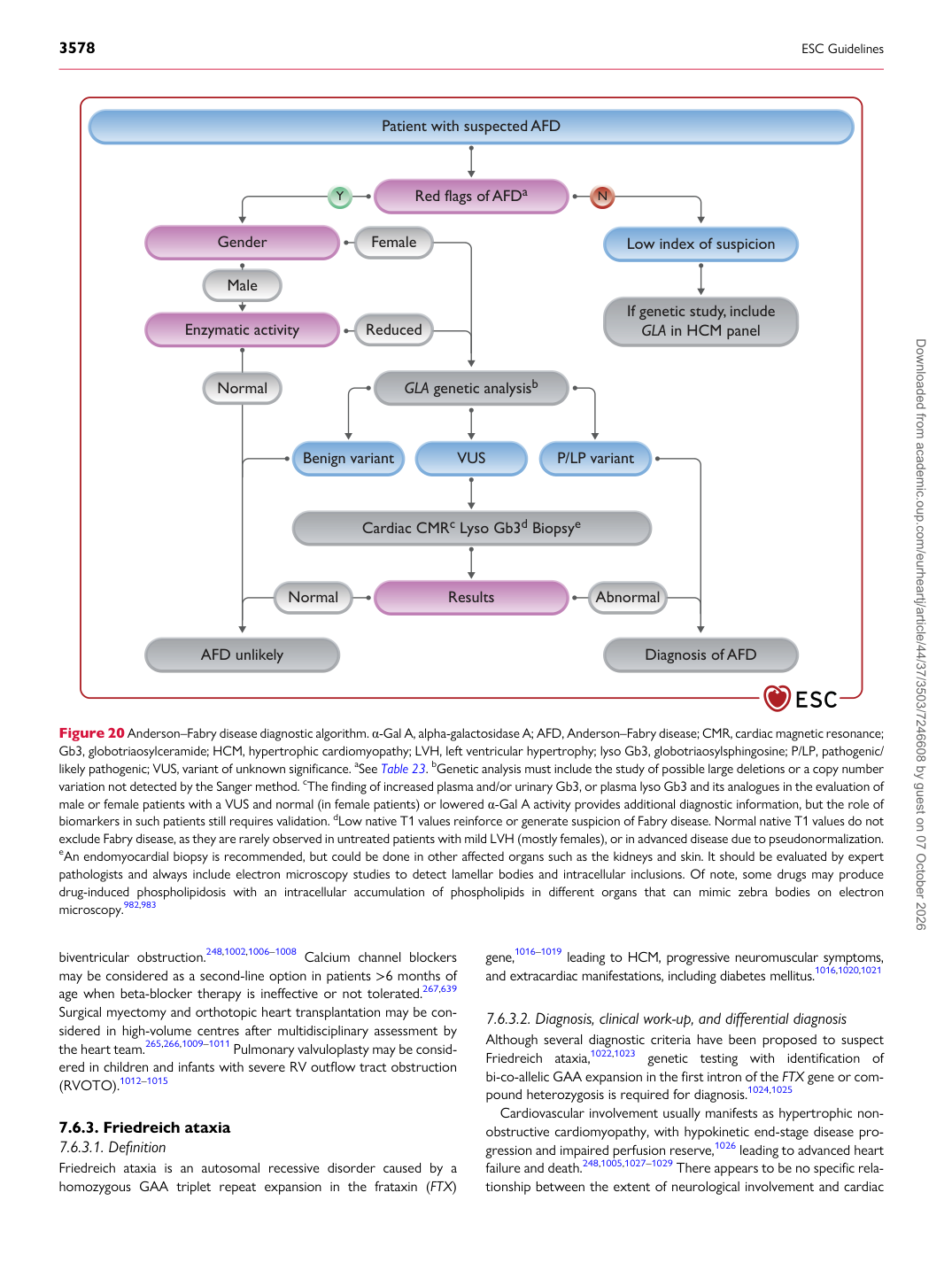
Figure 20 Anderson–Fabry disease diagnostic algorithm. α-Gal A, alpha-galactosidase A; AFD, Anderson–Fabry disease; CMR, cardiac magnetic resonance; Gb3, globotriaosylceramide; HCM, hypertrophic cardiomyopathy; LVH, left ventricular hypertrophy; lyso Gb3, globotriaosylsphingosine; P/LP, pathogenic/ likely pathogenic; VUS, variant of unknown significance. aSee Table 23. bGenetic analysis must include the study of possible large deletions or a copy number variation not detected by the Sanger method. cThe finding of increased plasma and/or urinary Gb3, or plasma lyso Gb3 and its analogues in the evaluation of male or female patients with a VUS and normal (in female patients) or lowered α-Gal A activity provides additional diagnostic information, but the role of biomarkers in such patients still requires validation. dLow native T1 values reinforce or generate suspicion of Fabry disease. Normal native T1 values do not exclude Fabry disease, as they are rarely observed in untreated patients with mild LVH (mostly females), or in advanced disease due to pseudonormalization. eAn endomyocardial biopsy is recommended, but could be done in other affected organs such as the kidneys and skin. It should be evaluated by expert pathologists and always include electron microscopy studies to detect lamellar bodies and intracellular inclusions. Of note, some drugs may produce drug-induced phospholipidosis with an intracellular accumulation of phospholipids in different organs that can mimic zebra bodies on electron microscopy.982,983 Downloaded from academic.oup.com/eurheartj/article/44/37/3503/7246608 by guest on 07 October 2026