p.70 抽出テキスト
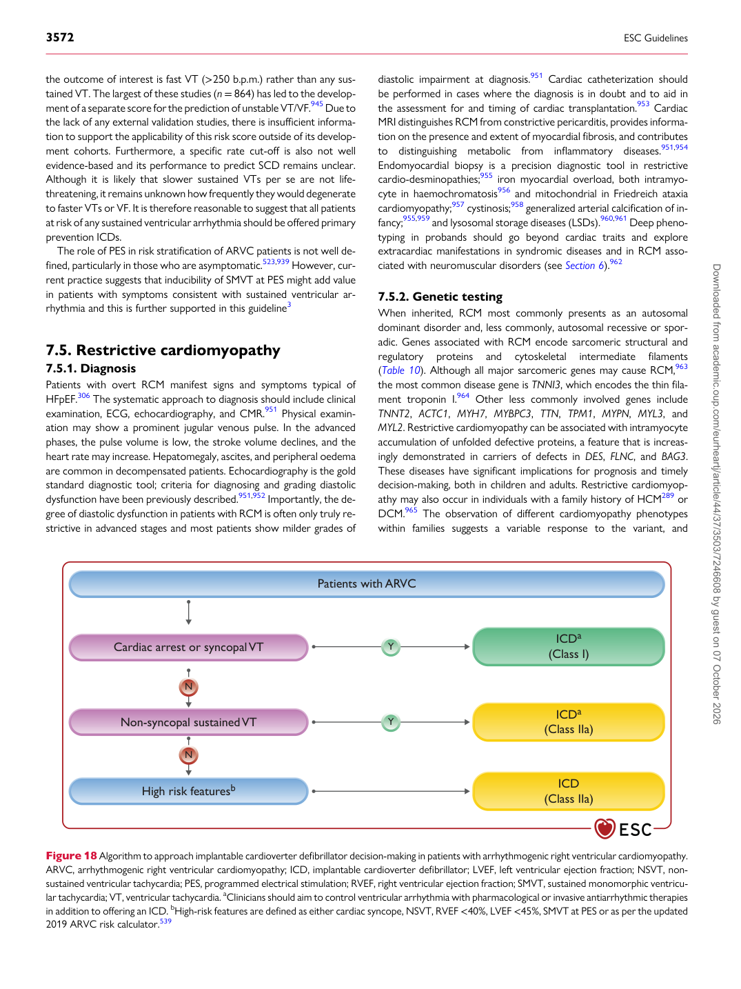
the outcome of interest is fast VT (>250 b.p.m.) rather than any sustained VT. The largest of these studies (n = 864) has led to the development of a separate score for the prediction of unstable VT/VF.945 Due to the lack of any external validation studies, there is insufficient information to support the applicability of this risk score outside of its development cohorts. Furthermore, a specific rate cut-off is also not well evidence-based and its performance to predict SCD remains unclear. Although it is likely that slower sustained VTs per se are not lifethreatening, it remains unknown how frequently they would degenerate to faster VTs or VF. It is therefore reasonable to suggest that all patients at risk of any sustained ventricular arrhythmia should be offered primary prevention ICDs. The role of PES in risk stratification of ARVC patients is not well defined, particularly in those who are asymptomatic.523,939 However, current practice suggests that inducibility of SMVT at PES might add value in patients with symptoms consistent with sustained ventricular arrhythmia and this is further supported in this guideline3

7.5 Restrictive cardiomyopathy

7.5.1. Diagnosis Patients with overt RCM manifest signs and symptoms typical of HFpEF.306 The systematic approach to diagnosis should include clinical examination, ECG, echocardiography, and CMR.951 Physical examination may show a prominent jugular venous pulse. In the advanced phases, the pulse volume is low, the stroke volume declines, and the heart rate may increase. Hepatomegaly, ascites, and peripheral oedema are common in decompensated patients. Echocardiography is the gold standard diagnostic tool; criteria for diagnosing and grading diastolic dysfunction have been previously described.951,952 Importantly, the degree of diastolic dysfunction in patients with RCM is often only truly restrictive in advanced stages and most patients show milder grades of diastolic impairment at diagnosis.951 Cardiac catheterization should be performed in cases where the diagnosis is in doubt and to aid in the assessment for and timing of cardiac transplantation.953 Cardiac MRI distinguishes RCM from constrictive pericarditis, provides information on the presence and extent of myocardial fibrosis, and contributes to distinguishing metabolic from inflammatory diseases.951,954

Endomyocardial biopsy is a precision diagnostic tool in restrictive cardio-desminopathies;955 iron myocardial overload, both intramyocyte in haemochromatosis956 and mitochondrial in Friedreich ataxia cardiomyopathy;957 cystinosis;958 generalized arterial calcification of infancy;955,959 and lysosomal storage diseases (LSDs).960,961 Deep phenotyping in probands should go beyond cardiac traits and explore extracardiac manifestations in syndromic diseases and in RCM associated with neuromuscular disorders (see Section 6).962

7.5.2 Genetic testing

When inherited, RCM most commonly presents as an autosomal dominant disorder and, less commonly, autosomal recessive or sporadic. Genes associated with RCM encode sarcomeric structural and regulatory proteins and cytoskeletal intermediate filaments (Table 10). Although all major sarcomeric genes may cause RCM,963 the most common disease gene is TNNI3, which encodes the thin filament troponin I.964 Other less commonly involved genes include TNNT2, ACTC1, MYH7, MYBPC3, TTN, TPM1, MYPN, MYL3, and MYL2. Restrictive cardiomyopathy can be associated with intramyocyte accumulation of unfolded defective proteins, a feature that is increasingly demonstrated in carriers of defects in DES, FLNC, and BAG3. These diseases have significant implications for prognosis and timely decision-making, both in children and adults. Restrictive cardiomyopathy may also occur in individuals with a family history of HCM289 or DCM.965 The observation of different cardiomyopathy phenotypes within families suggests a variable response to the variant, and

Cardiac arrest or syncopal VT

Non-syncopal sustained VT ICDa

(Class I)

ICDa (Class IIa)

N

N Patients with ARVC

Y

Y

High risk featuresb ICD (Class IIa)

Figure 18 Algorithm to approach implantable cardioverter defibrillator decision-making in patients with arrhythmogenic right ventricular cardiomyopathy. ARVC, arrhythmogenic right ventricular cardiomyopathy; ICD, implantable cardioverter defibrillator; LVEF, left ventricular ejection fraction; NSVT, nonsustained ventricular tachycardia; PES, programmed electrical stimulation; RVEF, right ventricular ejection fraction; SMVT, sustained monomorphic ventricular tachycardia; VT, ventricular tachycardia. aClinicians should aim to control ventricular arrhythmia with pharmacological or invasive antiarrhythmic therapies in addition to offering an ICD. bHigh-risk features are defined as either cardiac syncope, NSVT, RVEF <40%, LVEF <45%, SMVT at PES or as per the updated 2019 ARVC risk calculator.539 Downloaded from academic.oup.com/eurheartj/article/44/37/3503/7246608 by guest on 07 October 2026