p.68 抽出テキスト
7.4.1.8 Arrhythmogenic right ventricular cardiomyopathy

phenocopies In suspicion of ARVC, a systematic approach to investigation of phenocopies should be undertaken. Differential diagnosis in patients with suspected ARVC includes myocarditis, sarcoidosis, RV infarction, DCM, Chagas disease, pulmonary hypertension, and CHD with volume overload (such as Ebstein anomaly, atrial septal defect, and partial anomalous venous return, left-to-right shunt, and pericardial agenesis).909,910 Disease phenocopies also include non-structural diseases. In fact, one of the main diagnostic dilemmas is to distinguish ARVC from idiopathic RV outflow tract VT, since the latter is usually benign.4 The idiopathic nature of VT is supported by the absence of family history, a normal basal 12-lead ECG, a normal ventricular structure by cardiac imaging and electroanatomic mapping, a single VT morphology, and the noninducibility at programmed ventricular stimulation. In highly trained competitive athletes, differential diagnosis with physiological adaptation to training needs to be considered.911 Right ventricular enlargement, ECG abnormalities, and arrhythmias reflect the increased haemodynamic load during exercise. While global RV systolic dysfunction and/or RWMAs, such as bulgings or aneurysms, are more in keeping with ARVC, the absence of overt structural changes of the right ventricle, frequent VEs, or inverted T waves in pre-cordial leads all support a benign nature (so-called athlete’s heart).72,912,913

7.4.2 Genetic testing and family screening

The genes underlying ARVC mainly encode proteins of the cardiac desmosome: plakophilin-2 (PKP2), desmoplakin (DSP), desmoglein-2 (DSG2), desmocollin-2 (DSC2), and plakoglobin (JUP). In addition to desmosomal genes, P/LP variants have also been described in other genes, including DES,914 TMEM43,915 and PLN.190,882 Pathogenic or likely pathogenic variants can be identified in up to 60% of patients with a diagnosis of ARVC.230 Given the diagnostic importance of genetic testing in ARVC, it is important that genetic variants are frequently reappraised in terms of their pathogenicity.916 The pattern of inheritance in the majority of ARVC families is autosomal dominant. The penetrance of the disease in genetic carriers is age, gender, and physical activity dependent.892,917 Recommendations for clinical screening, genetic counselling, and testing are described in Sections 6.8.3 and 6.11. Cardiac evaluation should be adapted to the particular risk of complications in the family. Evaluation every 1–2 years including ECG, ECHO, and Holter/ECG monitoring is generally recommended for relatives at risk of developing the disease. Cardiac magnetic resonance should be considered at the baseline evaluation.

7.4.3 Assessment of symptoms

Patients with ARVC commonly experience palpitations and can develop symptoms of heart failure, although this may occur many years after the appearance of the initial abnormalities. Assessment of symptoms in patients with cardiomyopathies is described in Section 6.10.1.

7.4.4 Management

The aim of the clinical management of ARVC relies on the improvement of symptoms, the reduction of the pace of disease progression, and the prevention of complications. Recommendations for the pharmacological management of atrial arrhythmias and heart failure symptoms in patients with cardiomyopathies are described in Sections 6.10.2 and 6.10.3. 7.4.4.1 Antiarrhythmic therapy

Beta-blockers constitute the first option to reduce arrhythmic burden via a reduction in adrenergic tone, particularly on exercise. Titration to the maximal tolerated dose has been associated with an improvement in survival from major ventricular arrhythmias in retrospective observational studies.918 Amiodarone is often used when other beta-blockers fail to control arrhythmias.917,919,920 It should, however, be used with caution for the long-term management of ventricular arrhythmias, especially in young patients. Sotalol has been used for many years, but evidence regarding its efficacy remain limited and conflicting.921,922 Flecainide should be considered when single agent treatment has failed to control arrhythmia-related symptoms in patients with ARVC or when autonomic side effects limit the use of beta-blockers.923,924 Experience with other antiarrhythmics (dofetilide, ranolazine) is limited to very small case series.919,923 A proportion of patients require invasive arrhythmic procedures and/or ICD implantation. Complex endocardial and/or epicardial approach guided by three-dimensional (3D) electroanatomical mapping can be recommended but with a high recurrence rate (30–50% in experienced centres).919,925–927 Sympathetic denervation has also been used.928 Such procedures do not confer adequate protection against SCD, but may be very useful in reducing the VT burden and the risk of electrical storm.917 Discontinuation of intense physical exercise has shown a potential to slow the pace of disease progression and reduce the ventricular arrhythmia burden.917,919

7.4.5 Sudden cardiac death prevention in

arrhythmogenic right ventricular cardiomyopathy Arrhythmogenic right ventricular cardiomyopathy is characterized by its high propensity for ventricular arrhythmias and SCD.919 Although estimated to be a rare disease, it has been consistently reported as one of

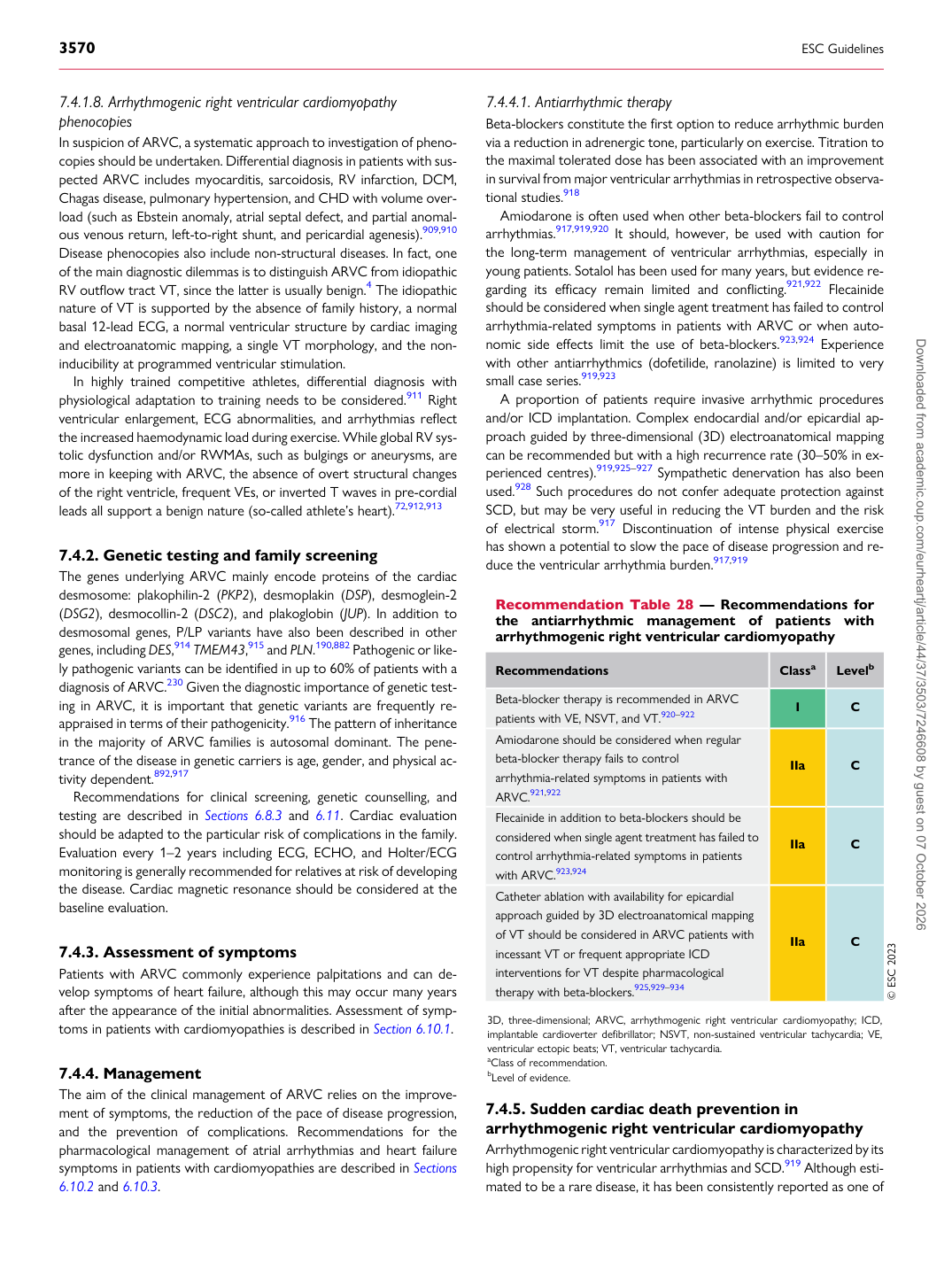
Recommendation Table 28 — Recommendations for the antiarrhythmic management of patients with arrhythmogenic right ventricular cardiomyopathy

Recommendations Classa Levelb

Beta-blocker therapy is recommended in ARVC patients with VE, NSVT, and VT.920–922 I C Amiodarone should be considered when regular beta-blocker therapy fails to control arrhythmia-related symptoms in patients with ARVC.921,922 IIa C Flecainide in addition to beta-blockers should be considered when single agent treatment has failed to control arrhythmia-related symptoms in patients with ARVC.923,924 IIa C Catheter ablation with availability for epicardial approach guided by 3D electroanatomical mapping of VT should be considered in ARVC patients with incessant VT or frequent appropriate ICD interventions for VT despite pharmacological therapy with beta-blockers.925,929–934 IIa C © ESC 2023

3D, three-dimensional; ARVC, arrhythmogenic right ventricular cardiomyopathy; ICD, implantable cardioverter defibrillator; NSVT, non-sustained ventricular tachycardia; VE, ventricular ectopic beats; VT, ventricular tachycardia. aClass of recommendation. bLevel of evidence. Downloaded from academic.oup.com/eurheartj/article/44/37/3503/7246608 by guest on 07 October 2026