p.67 抽出テキスト
7.4 Arrhythmogenic right ventricular

cardiomyopathy 7.4.1. Diagnosis 7.4.1.1. Index case Arrhythmogenic right ventricular cardiomyopathy (ARVC) is characterized structurally by a progressive myocardial atrophy with fibro-fatty replacement of the RV myocardium.890 Lesions can also be present in the LV myocardium; predominant LV disease can coexist in the same family. Arrhythmogenic right ventricular cardiomyopathy usually manifests in the second to fourth decade of life.891 Men are affected more frequently than women and an age-related penetrance has been demonstrated, with high clinical and genetic variability. An ARVC diagnosis should be suspected in adolescents or young adults with palpitations, syncope, or aborted sudden death. Frequent VEs or VT of LBBB morphology are among the most common clinical presentation. The presence of right pre-cordial TWI (V1–V3) in routine ECG testing should also be suspected for ARVC.10,892 Less common ECG changes include low QRS voltages in the peripheral leads and terminal activation delay in the right pre-cordial leads.893 Right ventricular dilatation on 2D echocardiography is also a frequent reason for patient referral. Less common presentations are RV or biventricular heart failure that can mimic DCM or NDLVC.894 Patients with multiple variants are thought to develop a more severe phenotype, and patients with a DSP or DSG2 variant are more prone to develop heart failure.895,896 In children and young adults, syncope, palpitations, and ventricular arrhythmias are also the usual presenting symptoms.897 However, chest pain, dynamic ST-T wave changes on basal 12-lead ECG, and myocardial enzymes release in the setting of normal coronary arteries are often reported, requiring differential diagnosis with myocarditis or acute myocardial infarction.898

7.4.1.2 Relatives

Clinical testing in relatives often reveals non-diagnostic abnormalities. In this context, the presence of RV systolic global or regional dysfunction, or additional electrocardiographic abnormalities (e.g. repolarization abnormalities, prolonged terminal activation duration, low QRS voltages, frequent ventricular extrasystoles [>500 per 24 h], or NSVT) in a firstdegree relative of an individual with ARVC (or a first-degree relative with autopsy-proven ARVC) is highly suggestive of ARVC and warrants close follow-up.

7.4.1.3 Diagnostic work-up

The key elements of the diagnostic work-up for all patients with ARVC are defined by the diagnostic criteria used for the identification of affected individuals. The revised Task Force criteria for the diagnosis of ARVC published by Marcus et al. in 2010 have been used for the diagnosis of ARVC for more than a decade.10 More recently, the Padua criteria have offered an updated iteration to include LV involvement but are yet to be externally validated.5 Key elements of the diagnostic workup include ECG, Holter monitoring, cardiac imaging, genetic testing, and, in specific circumstances, EMB.4,10,892 Additional laboratory tests, exercise testing, and cardiac catheterization should also be considered, as detailed in Section 6.

7.4.1.4 Electrocardiography and Holter monitoring

Abnormalities of the repolarization and depolarization as well as arrhythmias are key to the diagnosis of ARVC.5 The diagnostic utility of late potentials on signal-averaged electrocardiogram (SAECG) has been challenged in patients with ARVC for showing poor sensitivity and specificity.5,899 It has been noted that epsilon waves are frequently overdiagnosed and that there is poor agreement even between experts regarding their presence.900 Furthermore, it has been demonstrated that they occur in the presence of severe structural disease and thus add little to the diagnosis.900,901 Therefore, epsilon waves and SAECG should be utilized for diagnostic purposes with caution.

7.4.1.5 Echocardiography and cardiac magnetic resonance

A comprehensive cardiac imaging assessment is recommended for all ARVC patients.71,73 Structural and functional alterations assessed by echocardiography and CMR are key to ARVC diagnosis.10 The key feature is the presence of wall motional abnormalities such as RV akinesia, dyskinesia, or bulging, and the determinant of the diagnostic performance is the level of RV dilatation or dysfunction (major and minor criteria). Cardiac magnetic resonance should be considered the first-line test for assessment of the RV functional structural abnormalities criterion as it has demonstrated superior sensitivity.10 Contrast-enhanced CMR is the only tool allowing the detection of LV involvement which remains otherwise underestimated by applying the 2010 Task Force Criteria. Tissue characterization by CMR or indirectly by electroanatomical voltage mapping may show signs of fibro-fatty replacement that can be present in either ventricle.889,903,904

7.4.1.6 Endomyocardial biopsy

Differential diagnosis in patients with suspected ARVC includes inflammatory processes affecting the right ventricle such as myocarditis and sarcoidosis. In some instances, especially when dealing with probands with a sporadic form, EMB may be helpful to rule out myocarditis and sarcoidosis.72,892,905 Endomyocardial biopsy can also be useful in selected patients in whom non-invasive assessment is inconclusive.4,72 Electroanatomic voltage mapping-guided EMB may be considered in selected cases, particularly in case of negative CMR.906

7.4.1.7 Nuclear medicine

Measurement of 18F-FDG uptake using PET, with focal or focal-on-diffuse FDG uptake patterns, can be useful in suspected cardiac sarcoidosis.849 However, it has been demonstrated that patients with ARVC can also show myocardial 18F-FDG-PET uptake.888,907 Therefore, there is a limited role for radionuclide imaging in ARVC unless there is concomitant abnormal 18F-FDG-PET uptake in extracardiac tissues, or other clinical features suggestive of cardiac sarcoidosis.904,908

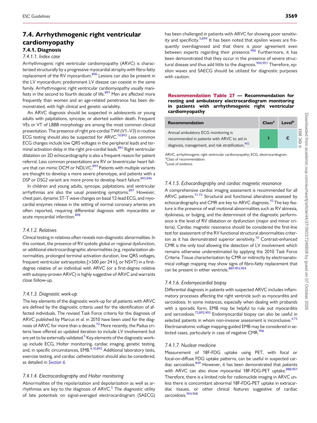
Recommendation Table 27 — Recommendation for resting and ambulatory electrocardiogram monitoring in patients with arrhythmogenic right ventricular cardiomyopathy

Recommendation Classa Levelb

Annual ambulatory ECG monitoring is recommended in patients with ARVC to aid in diagnosis, management, and risk stratification.902 I C © ESC 2023

ARVC, arrhythmogenic right ventricular cardiomyopathy; ECG, electrocardiogram. aClass of recommendation. bLevel of evidence.

ESC Guidelines                                                                                                                                                                                          3569

Downloaded from academic.oup.com/eurheartj/article/44/37/3503/7246608 by guest on 07 October 2026