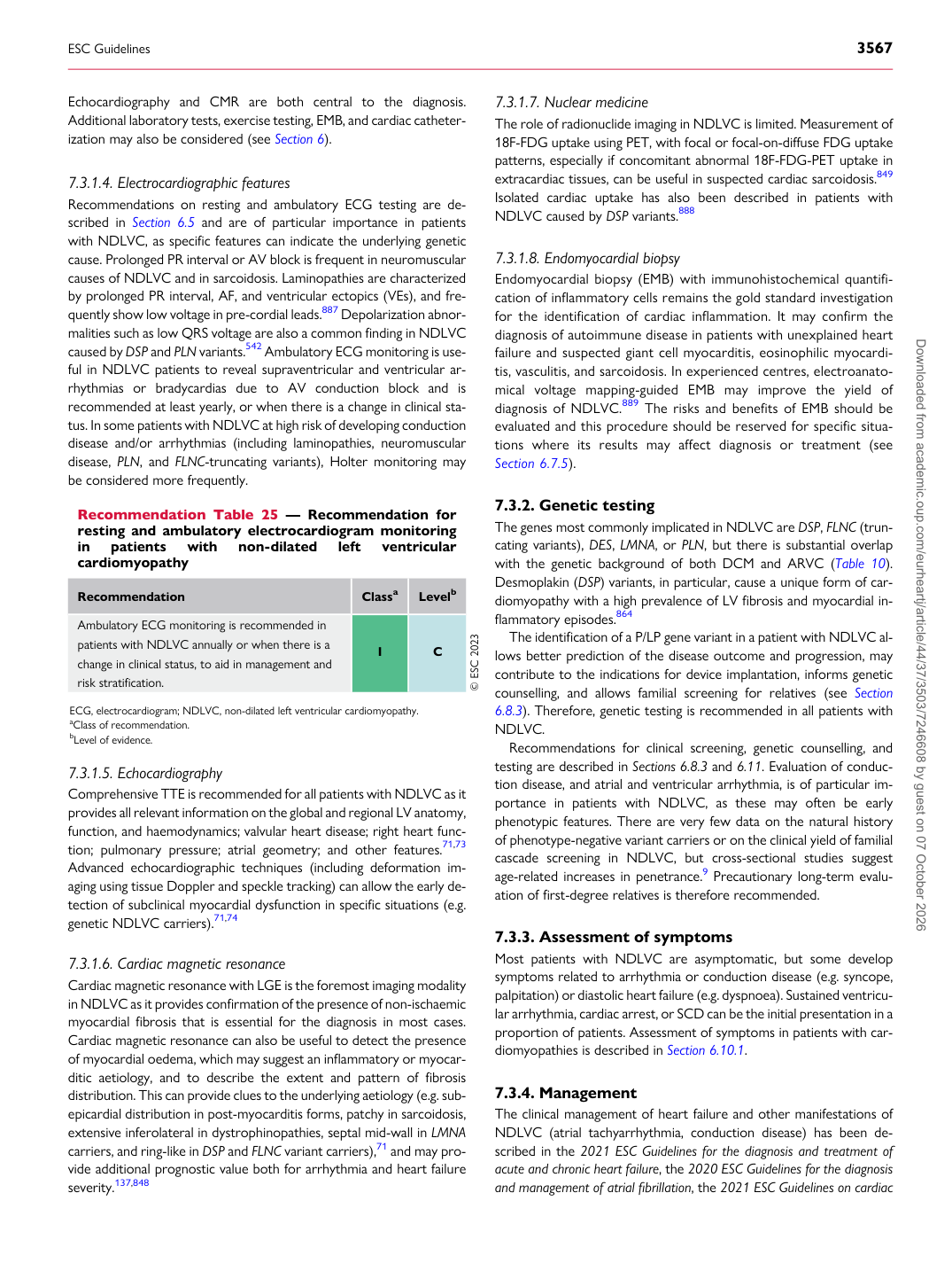
p.65 抽出テキスト
Echocardiography and CMR are both central to the diagnosis. Additional laboratory tests, exercise testing, EMB, and cardiac catheterization may also be considered (see Section 6).

7.3.1.4 Electrocardiographic features

Recommendations on resting and ambulatory ECG testing are described in Section 6.5 and are of particular importance in patients with NDLVC, as specific features can indicate the underlying genetic cause. Prolonged PR interval or AV block is frequent in neuromuscular causes of NDLVC and in sarcoidosis. Laminopathies are characterized by prolonged PR interval, AF, and ventricular ectopics (VEs), and frequently show low voltage in pre-cordial leads.887 Depolarization abnormalities such as low QRS voltage are also a common finding in NDLVC caused by DSP and PLN variants.542 Ambulatory ECG monitoring is useful in NDLVC patients to reveal supraventricular and ventricular arrhythmias or bradycardias due to AV conduction block and is recommended at least yearly, or when there is a change in clinical status. In some patients with NDLVC at high risk of developing conduction disease and/or arrhythmias (including laminopathies, neuromuscular disease, PLN, and FLNC-truncating variants), Holter monitoring may be considered more frequently.

7.3.1.5 Echocardiography

Comprehensive TTE is recommended for all patients with NDLVC as it provides all relevant information on the global and regional LV anatomy, function, and haemodynamics; valvular heart disease; right heart function; pulmonary pressure; atrial geometry; and other features.71,73 Advanced echocardiographic techniques (including deformation imaging using tissue Doppler and speckle tracking) can allow the early detection of subclinical myocardial dysfunction in specific situations (e.g. genetic NDLVC carriers).71,74

7.3.1.6 Cardiac magnetic resonance

Cardiac magnetic resonance with LGE is the foremost imaging modality in NDLVC as it provides confirmation of the presence of non-ischaemic myocardial fibrosis that is essential for the diagnosis in most cases. Cardiac magnetic resonance can also be useful to detect the presence of myocardial oedema, which may suggest an inflammatory or myocarditic aetiology, and to describe the extent and pattern of fibrosis distribution. This can provide clues to the underlying aetiology (e.g. subepicardial distribution in post-myocarditis forms, patchy in sarcoidosis, extensive inferolateral in dystrophinopathies, septal mid-wall in LMNA carriers, and ring-like in DSP and FLNC variant carriers),71 and may provide additional prognostic value both for arrhythmia and heart failure severity.137,848 7.3.1.7 Nuclear medicine

The role of radionuclide imaging in NDLVC is limited. Measurement of 18F-FDG uptake using PET, with focal or focal-on-diffuse FDG uptake patterns, especially if concomitant abnormal 18F-FDG-PET uptake in extracardiac tissues, can be useful in suspected cardiac sarcoidosis.849 Isolated cardiac uptake has also been described in patients with NDLVC caused by DSP variants.888

7.3.1.8 Endomyocardial biopsy

Endomyocardial biopsy (EMB) with immunohistochemical quantification of inflammatory cells remains the gold standard investigation for the identification of cardiac inflammation. It may confirm the diagnosis of autoimmune disease in patients with unexplained heart failure and suspected giant cell myocarditis, eosinophilic myocarditis, vasculitis, and sarcoidosis. In experienced centres, electroanatomical voltage mapping-guided EMB may improve the yield of diagnosis of NDLVC.889 The risks and benefits of EMB should be evaluated and this procedure should be reserved for specific situations where its results may affect diagnosis or treatment (see Section 6.7.5).

7.3.2 Genetic testing

The genes most commonly implicated in NDLVC are DSP, FLNC (truncating variants), DES, LMNA, or PLN, but there is substantial overlap with the genetic background of both DCM and ARVC (Table 10). Desmoplakin (DSP) variants, in particular, cause a unique form of cardiomyopathy with a high prevalence of LV fibrosis and myocardial inflammatory episodes.864 The identification of a P/LP gene variant in a patient with NDLVC allows better prediction of the disease outcome and progression, may contribute to the indications for device implantation, informs genetic counselling, and allows familial screening for relatives (see Section 6.8.3). Therefore, genetic testing is recommended in all patients with NDLVC. Recommendations for clinical screening, genetic counselling, and testing are described in Sections 6.8.3 and 6.11. Evaluation of conduction disease, and atrial and ventricular arrhythmia, is of particular importance in patients with NDLVC, as these may often be early phenotypic features. There are very few data on the natural history of phenotype-negative variant carriers or on the clinical yield of familial cascade screening in NDLVC, but cross-sectional studies suggest age-related increases in penetrance.9 Precautionary long-term evaluation of first-degree relatives is therefore recommended.

7.3.3 Assessment of symptoms

Most patients with NDLVC are asymptomatic, but some develop symptoms related to arrhythmia or conduction disease (e.g. syncope, palpitation) or diastolic heart failure (e.g. dyspnoea). Sustained ventricular arrhythmia, cardiac arrest, or SCD can be the initial presentation in a proportion of patients. Assessment of symptoms in patients with cardiomyopathies is described in Section 6.10.1.

7.3.4 Management

The clinical management of heart failure and other manifestations of NDLVC (atrial tachyarrhythmia, conduction disease) has been described in the 2021 ESC Guidelines for the diagnosis and treatment of acute and chronic heart failure, the 2020 ESC Guidelines for the diagnosis and management of atrial fibrillation, the 2021 ESC Guidelines on cardiac

Recommendation Table 25 — Recommendation for resting and ambulatory electrocardiogram monitoring in patients with non-dilated left ventricular cardiomyopathy

Recommendation Classa Levelb

Ambulatory ECG monitoring is recommended in patients with NDLVC annually or when there is a change in clinical status, to aid in management and risk stratification. I C © ESC 2023

ECG, electrocardiogram; NDLVC, non-dilated left ventricular cardiomyopathy. aClass of recommendation. bLevel of evidence.

ESC Guidelines                                                                                                                                                                                          3567

Downloaded from academic.oup.com/eurheartj/article/44/37/3503/7246608 by guest on 07 October 2026