p.61 抽出テキスト
7.2.1.5 Cardiac magnetic resonance

Cardiac magnetic resonance provides additional information on tissue characterization in patients with DCM, including the presence of myocardial oedema, which may suggest a myocarditic or inflammatory cause, and LGE, to determine the presence and extent of fibrosis, as well as its distribution, which may allow exclusion of myocardial infarction and also point towards specific aetiologies (e.g. subepicardial distribution in post-myocarditis forms, patchy in sarcoidosis, extensive inferolateral in dystrophinopathies, septal mid-wall in LMNA carriers, and ring-like in DSP and FLNC-truncating variant carriers) (see Section 7.3).71,847 Late gadolinium enhancement distribution and extent hold prognostic value both for arrhythmia and heart failure severity.137,848 Dedicated T2* sequences describe myocardial iron deposition, which is useful for the diagnosis of haemochromatosis.71

7.2.1.6 Nuclear medicine

There is a limited role for radionuclide imaging in DCM. Measurement of 18F-fluorodeoxyglucose (18F-FDG) uptake using PET, with focal or focal-on-diffuse FDG uptake patterns especially if there is concomitant abnormal 18F-FDG-PET uptake in extracardiac tissues, can be useful in suspected cardiac sarcoidosis.849

7.2.2 Genetic testing and family screening

The aetiology of DCM is highly heterogeneous and includes inherited (genetic/familial) and acquired causes.9,545,850,851 Direct causes of DCM include pathogenic gene variants, toxins, auto-immunity, infections, storage diseases, and tachyarrhythmias. Monogenic gene variants causing DCM are highly heterogeneous, implicating many genes and diverse pathways. Moreover, only 30–40% of DCM cases are attributable to pathogenic rare variants, with a substantial polygenic/common variant contribution in this population. Furthermore, disease modifiers can play a role in the acceleration of the DCM phenotype.7,9,850 This includes conditions that may aggravate or trigger DCM, including epigenetic factors and acquired modifiers, such as pregnancy, hypertension, excessive alcohol use, and other toxins.42–44 It is important to consider the interplay between genetic and acquired causes during the diagnostic work-up. Identification of an acquired cause does not exclude an underlying causative gene variant, whereas the latter may require an additional acquired cause and/or disease modifier to manifest. Within the genes that can cause DCM, there are genes robustly associated with classical DCM that have been recently curated,189 and also others classically associated with ARVC but that very commonly can present with LV dilatation and predominantly LV dysfunction. Moreover, genes described in the context of hypertrabeculation/LVNC (e.g. NKX2.5 and PRDM16), or that can cause DCM with or without skeletal involvement (such as DMD or EMD), should also be considered DCM-associated genes and examined, particularly if phenotype is concordant. The most common genetic and acquired causes of DCM are shown in Table 10 and Table 20. Detailed lists of causes of DCM have been previously published.9,852

7.2.2.1 Genetic testing

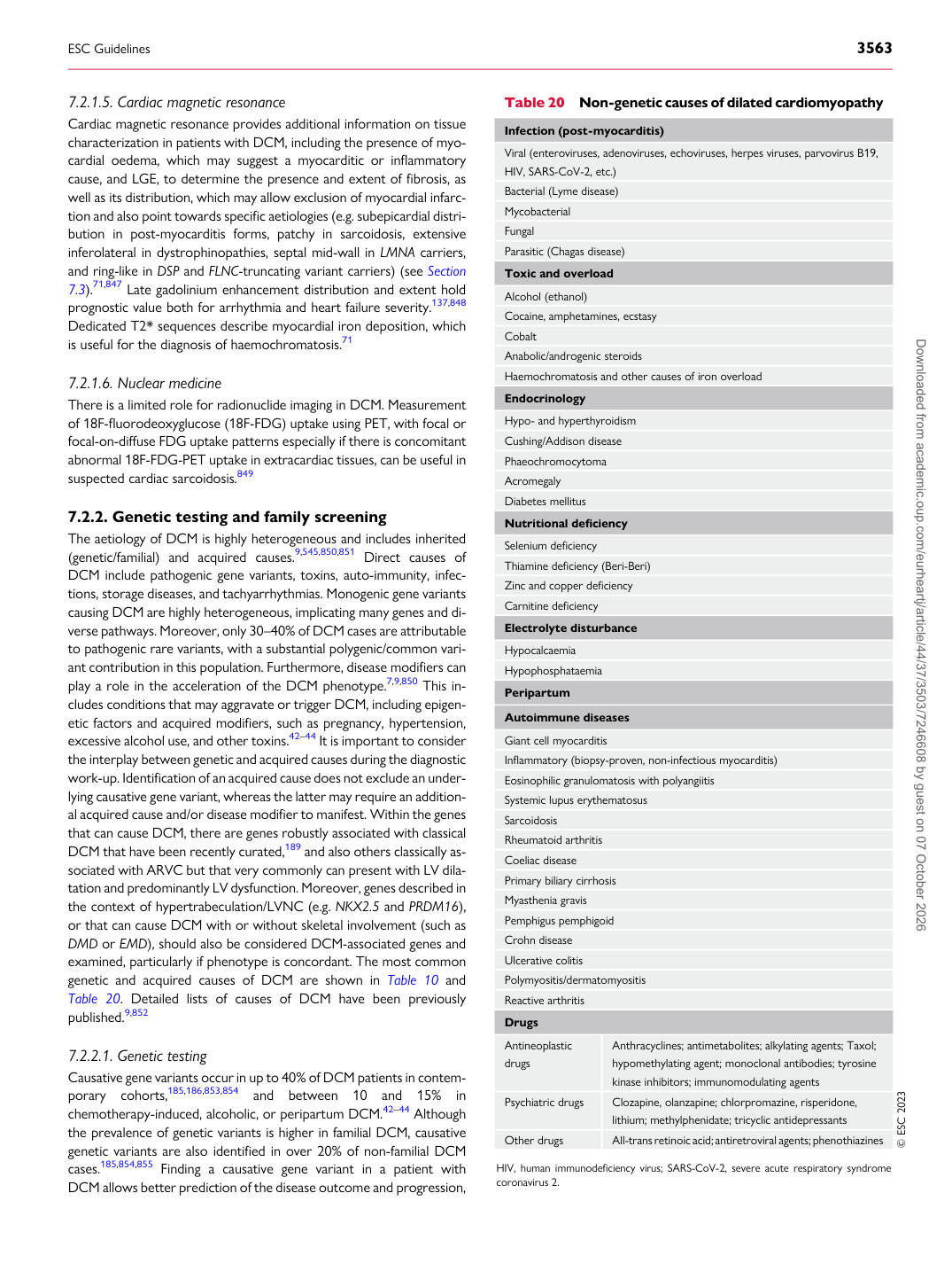
Causative gene variants occur in up to 40% of DCM patients in contemporary cohorts,185,186,853,854 and between 10 and 15% in chemotherapy-induced, alcoholic, or peripartum DCM.42–44 Although the prevalence of genetic variants is higher in familial DCM, causative genetic variants are also identified in over 20% of non-familial DCM cases.185,854,855 Finding a causative gene variant in a patient with DCM allows better prediction of the disease outcome and progression, Table 20 Non-genetic causes of dilated cardiomyopathy

Infection (post-myocarditis) Viral (enteroviruses, adenoviruses, echoviruses, herpes viruses, parvovirus B19, HIV, SARS-CoV-2, etc.) Bacterial (Lyme disease) Mycobacterial Fungal Parasitic (Chagas disease) Toxic and overload Alcohol (ethanol) Cocaine, amphetamines, ecstasy Cobalt Anabolic/androgenic steroids Haemochromatosis and other causes of iron overload Endocrinology Hypo- and hyperthyroidism Cushing/Addison disease Phaeochromocytoma Acromegaly Diabetes mellitus Nutritional deficiency Selenium deficiency Thiamine deficiency (Beri-Beri) Zinc and copper deficiency Carnitine deficiency Electrolyte disturbance Hypocalcaemia Hypophosphataemia Peripartum Autoimmune diseases Giant cell myocarditis Inflammatory (biopsy-proven, non-infectious myocarditis) Eosinophilic granulomatosis with polyangiitis Systemic lupus erythematosus Sarcoidosis Rheumatoid arthritis Coeliac disease Primary biliary cirrhosis Myasthenia gravis Pemphigus pemphigoid Crohn disease Ulcerative colitis Polymyositis/dermatomyositis Reactive arthritis Drugs Antineoplastic drugs Anthracyclines; antimetabolites; alkylating agents; Taxol;

hypomethylating agent; monoclonal antibodies; tyrosine kinase inhibitors; immunomodulating agents

Psychiatric drugs Clozapine, olanzapine; chlorpromazine, risperidone,

lithium; methylphenidate; tricyclic antidepressants

Other drugs All-trans retinoic acid; antiretroviral agents; phenothiazines © ESC 2023

HIV, human immunodeficiency virus; SARS-CoV-2, severe acute respiratory syndrome coronavirus 2.

ESC Guidelines                                                                                                                                                                                          3563

Downloaded from academic.oup.com/eurheartj/article/44/37/3503/7246608 by guest on 07 October 2026