p.60 抽出テキスト
7.2 Dilated cardiomyopathy

7.2.1. Diagnosis 7.2.1.1. Index case Dilated cardiomyopathy is defined by the presence of LV dilatation and systolic dysfunction unexplained solely by abnormal loading conditions or CAD. Left ventricular dilatation is defined by LV end-diastolic dimensions or volumes >2 z-scores above population mean values corrected for body size, sex, and/or age. For adults this represents an LV enddiastolic diameter >58 mm in males and >52 in females and an LVEDV index of ≥75 mL/m2 in males and ≥62 mL/m2 in females by ECHO.9,845,846 Left ventricular global systolic dysfunction is defined by LVEF <50%.9

7.2.1.2 Relatives

Clinical testing in relatives often reveals mild non-diagnostic abnormalities that overlap with normal variation or mimic changes seen in other more common diseases such as hypertension and obesity. In this context, the presence of isolated LV dilatation with preserved systolic function or in the presence of a familial causative variant is sufficient for a diagnosis of DCM in a relative. Additional electrocardiographic or imaging abnormalities in the context of a family history of DCM are suggestive of disease and warrant close follow-up.9,75,817 In the absence of conclusive genetic information in a family, DCM is considered familial if: (i) one or more first- or second-degree relatives have DCM; or (ii) when an otherwise unexplained SCD has occurred in a first-degree relative at any age with an established diagnosis of DCM.

7.2.1.3 Diagnostic work-up

The key elements of the diagnostic work-up for all patients with DCM are described in Section 6 and include clinical and family history, laboratory tests, ECG, Holter monitoring, cardiac imaging, and genetic testing. Echocardiography is central for the diagnosis and CMR provides more detailed morphological and prognostic information. Additional laboratory tests, exercise testing, EMB, cardiac CT, and cardiac catheterization should also be considered, as detailed in Section 6.

7.2.1.4 Echocardiography

Comprehensive TTE is recommended for all DCM patients as it provides all the relevant information on the global and regional LV anatomy, function and haemodynamics, valvular heart disease, right heart function, pulmonary pressure, atrial geometry, and associated features.71 Advanced echocardiographic techniques (tissue Doppler and speckle tracking deformation imaging) can allow the early detection of subclinical myocardial dysfunction in specific situations (e.g. genetic DCM carriers, recipients of known cardiotoxic chemotherapy).71,74 Contrast agents may be considered for better endocardial delineation, to better depict the presence of hypertrabeculation, or to exclude intraventricular thrombus. Transoesophageal echocardiography is rarely necessary except for when atrial thrombi are present in patients with AF, or for assessing valvular function and guiding transcatheter therapy in patients with concomitant secondary mitral or tricuspid regurgitation.

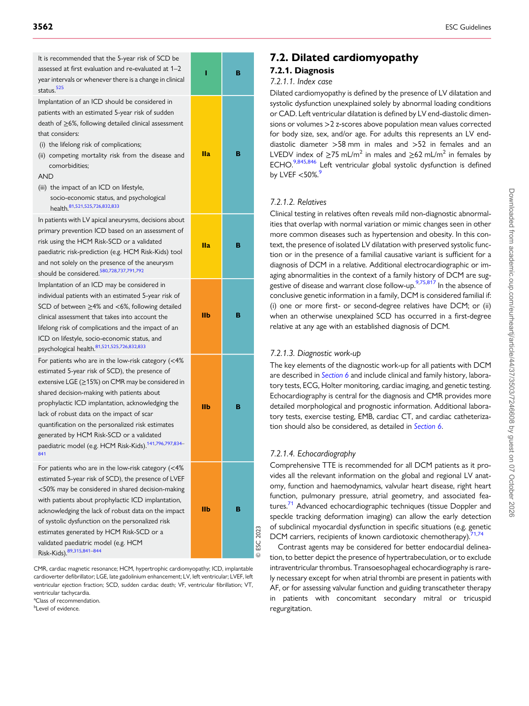
It is recommended that the 5-year risk of SCD be assessed at first evaluation and re-evaluated at 1–2 year intervals or whenever there is a change in clinical status.525 I B Implantation of an ICD should be considered in patients with an estimated 5-year risk of sudden death of ≥6%, following detailed clinical assessment that considers: (i) the lifelong risk of complications; (ii) competing mortality risk from the disease and comorbidities; AND (iii) the impact of an ICD on lifestyle,

socio-economic status, and psychological health.81,521,525,726,832,833 IIa B In patients with LV apical aneurysms, decisions about primary prevention ICD based on an assessment of risk using the HCM Risk-SCD or a validated paediatric risk-prediction (e.g. HCM Risk-Kids) tool and not solely on the presence of the aneurysm should be considered.580,728,737,791,792 IIa B Implantation of an ICD may be considered in individual patients with an estimated 5-year risk of SCD of between ≥4% and <6%, following detailed clinical assessment that takes into account the lifelong risk of complications and the impact of an ICD on lifestyle, socio-economic status, and psychological health.81,521,525,726,832,833 IIb B For patients who are in the low-risk category (<4% estimated 5-year risk of SCD), the presence of extensive LGE (≥15%) on CMR may be considered in shared decision-making with patients about prophylactic ICD implantation, acknowledging the lack of robust data on the impact of scar quantification on the personalized risk estimates generated by HCM Risk-SCD or a validated paediatric model (e.g. HCM Risk-Kids).141,796,797,834–

841 IIb B

For patients who are in the low-risk category (<4% estimated 5-year risk of SCD), the presence of LVEF <50% may be considered in shared decision-making with patients about prophylactic ICD implantation, acknowledging the lack of robust data on the impact of systolic dysfunction on the personalized risk estimates generated by HCM Risk-SCD or a validated paediatric model (e.g. HCM Risk-Kids).89,315,841–844 IIb B © ESC 2023

CMR, cardiac magnetic resonance; HCM, hypertrophic cardiomyopathy; ICD, implantable cardioverter defibrillator; LGE, late gadolinium enhancement; LV, left ventricular; LVEF, left ventricular ejection fraction; SCD, sudden cardiac death; VF, ventricular fibrillation; VT, ventricular tachycardia. aClass of recommendation. bLevel of evidence. Downloaded from academic.oup.com/eurheartj/article/44/37/3503/7246608 by guest on 07 October 2026