p.49 抽出テキスト
7.1.1.5 Nuclear imaging

The major clinical contribution of nuclear imaging in HCM is the detection of TTR-related cardiac amyloidosis (see Section 7.7). Recommendations on the utility of bone scintigraphy and cardiac CT are described in Section 6.7.4. 7.1.2 Genetic testing and family screening

In about half of cases, HCM is inherited as a Mendelian genetic trait. In such cases, the inheritance is primarily autosomal dominant, i.e. with a 50% risk of transmission to offspring.608 Apparently sporadic cases can have a monogenic cause, either because of incomplete penetrance of a

Table 18 Echocardiographic features that suggest specific aetiologies in hypertrophic cardiomyopathy

Finding Specific diseases to be considered

Increased interatrial septum thickness Amyloidosis Increased AV valve thickness Amyloidosis; Anderson–Fabry disease Increased RV free wall thickness Amyloidosis, myocarditis, Anderson–Fabry disease, Noonan syndrome, and related disorders Mild-to-moderate pericardial effusion Amyloidosis, myocarditis/myopericarditis Ground-glass appearance of ventricular myocardium on 2D echocardiography Amyloidosis Concentric LVH Glycogen storage disease, Anderson–Fabry disease, PRKAG2 variants, Friedreich ataxia Extreme concentric LVH (wall thickness ≥30 mm) Danon disease, Pompe disease Global LV hypokinesia (with or without LV dilatation) Mitochondrial disease, TTR-related amyloidosis, PRKAG2 variants, Danon disease, myocarditis, advanced sarcomeric HCM, Anderson–Fabry disease, Friedreich ataxia RVOTO Noonan syndrome and associated disorders Apical sparing pattern on longitudinal strain imaging Amyloidosis © ESC 2023

2D, two-dimensional; AV, atrioventricular; HCM, hypertrophic cardiomyopathy; LV, left ventricular; LVH, left ventricular hypertrophy; PRKAG2, protein kinase AMP-activated non-catalytic subunit gamma 2; RV, right ventricular; RVOTO, right ventricular outflow tract obstruction; TTR, transthyretin. Modified from Rapezzi et al.62

N

N

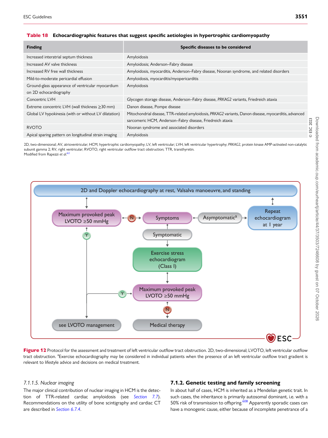
2D and Doppler echocardiography at rest,  Valsalva manoeuvre, and standing

Maximum provoked peak

LVOTO ≥50 mmHg

Maximum provoked peak

LVOTO ≥50 mmHg

Symptoms

Exercise stress echocardiogram

(Class I) Repeat echocardiogram

at 1 year

see LVOTO management Medical therapy

Y

Y Asymptomatica

Symptomatic

Figure 12 Protocol for the assessment and treatment of left ventricular outflow tract obstruction. 2D, two-dimensional; LVOTO, left ventricular outflow tract obstruction. aExercise echocardiography may be considered in individual patients when the presence of an left ventricular outflow tract gradient is relevant to lifestyle advice and decisions on medical treatment.

ESC Guidelines                                                                                                                                                                                          3551

Downloaded from academic.oup.com/eurheartj/article/44/37/3503/7246608 by guest on 07 October 2026