p.48 抽出テキスト
7.1.1.4 Cardiac magnetic resonance

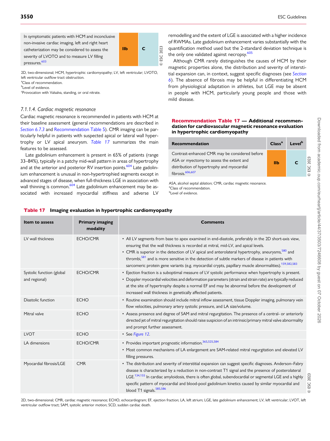
Cardiac magnetic resonance is recommended in patients with HCM at their baseline assessment (general recommendations are described in Section 6.7.3 and Recommendation Table 5). CMR imaging can be particularly helpful in patients with suspected apical or lateral wall hypertrophy or LV apical aneurysm. Table 17 summarizes the main features to be assessed. Late gadolinium enhancement is present in 65% of patients (range 33–84%), typically in a patchy mid-wall pattern in areas of hypertrophy and at the anterior and posterior RV insertion points.604 Late gadolinium enhancement is unusual in non-hypertrophied segments except in advanced stages of disease, when full-thickness LGE in association with wall thinning is common.604 Late gadolinium enhancement may be associated with increased myocardial stiffness and adverse LV remodelling and the extent of LGE is associated with a higher incidence of RWMAs. Late gadolinium enhancement varies substantially with the quantification method used but the 2-standard deviation technique is the only one validated against necropsy.605

Although CMR rarely distinguishes the causes of HCM by their magnetic properties alone, the distribution and severity of interstitial expansion can, in context, suggest specific diagnoses (see Section 6). The absence of fibrosis may be helpful in differentiating HCM from physiological adaptation in athletes, but LGE may be absent in people with HCM, particularly young people and those with mild disease.

Table 17 Imaging evaluation in hypertrophic cardiomyopathy

Item to assess Primary imaging

modality Comments

LV wall thickness ECHO/CMR • All LV segments from base to apex examined in end-diastole, preferably in the 2D short-axis view, ensuring that the wall thickness is recorded at mitral, mid-LV, and apical levels.

• CMR is superior in the detection of LV apical and anterolateral hypertrophy, aneurysms,580 and

thrombi,581 and is more sensitive in the detection of subtle markers of disease in patients with sarcomeric protein gene variants (e.g. myocardial crypts, papillary muscle abnormalities).159,582,583

Systolic function (global and regional) ECHO/CMR • Ejection fraction is a suboptimal measure of LV systolic performance when hypertrophy is present. • Doppler myocardial velocities and deformation parameters (strain and strain rate) are typically reduced

at the site of hypertrophy despite a normal EF and may be abnormal before the development of increased wall thickness in genetically affected patients.

Diastolic function ECHO • Routine examination should include mitral inflow assessment, tissue Doppler imaging, pulmonary vein

flow velocities, pulmonary artery systolic pressure, and LA size/volume.

Mitral valve ECHO • Assess presence and degree of SAM and mitral regurgitation. The presence of a central- or anteriorly

directed jet of mitral regurgitation should raise suspicion of an intrinsic/primary mitral valve abnormality and prompt further assessment.

LVOT ECHO • See Figure 12. LA dimensions ECHO/CMR • Provides important prognostic information.365,525,584

• Most common mechanisms of LA enlargement are SAM-related mitral regurgitation and elevated LV

filling pressures.

Myocardial fibrosis/LGE CMR • The distribution and severity of interstitial expansion can suggest specific diagnoses. Anderson–Fabry

disease is characterized by a reduction in non-contrast T1 signal and the presence of posterolateral LGE.134,155 In cardiac amyloidosis, there is often global, subendocardial or segmental LGE and a highly specific pattern of myocardial and blood-pool gadolinium kinetics caused by similar myocardial and blood T1 signals.585,586 © ESC 2023

2D, two-dimensional; CMR, cardiac magnetic resonance; ECHO, echocardiogram; EF, ejection fraction; LA, left atrium; LGE, late gadolinium enhancement; LV, left ventricular; LVOT, left ventricular outflow tract; SAM, systolic anterior motion; SCD, sudden cardiac death.

In symptomatic patients with HCM and inconclusive non-invasive cardiac imaging, left and right heart catheterization may be considered to assess the severity of LVOTO and to measure LV filling pressures.603 IIb C © ESC 2023

2D, two-dimensional; HCM, hypertrophic cardiomyopathy; LV, left ventricular; LVOTO, left ventricular outflow tract obstruction. aClass of recommendation. bLevel of evidence. cProvocation with Valsalva, standing, or oral nitrate.

Recommendation Table 17 — Additional recommendation for cardiovascular magnetic resonance evaluation in hypertrophic cardiomyopathy

Recommendation Classa Levelb

Contrast-enhanced CMR may be considered before ASA or myectomy to assess the extent and distribution of hypertrophy and myocardial fibrosis.606,607 IIb C © ESC 2023

ASA, alcohol septal ablation; CMR, cardiac magnetic resonance. aClass of recommendation. bLevel of evidence. Downloaded from academic.oup.com/eurheartj/article/44/37/3503/7246608 by guest on 07 October 2026