p.47 抽出テキスト
6.13 The patient pathway

The systematic, multiparametric approach to diagnosis and evaluation of patients with suspected cardiomyopathy described in this section allows clinicians to establish the presence of a cardiomyopathy and identify its aetiology and guides the management of symptoms and prevention of disease-related complications. While many of the aspects of clinical care and the accompanying recommendations are common to all cardiomyopathy phenotypes, achieving an aetiological diagnosis is key to delivering disease-specific management; this is discussed in detail in the subsequent sections of this guideline (see Section 7).

7 Specific cardiomyopathy

phenotypes

7.1 Hypertrophic cardiomyopathy

The 2014 ESC Guidelines on diagnosis and management of hypertrophic cardiomyopathy provide detailed recommendations on the assessment and management of patients with HCM.1 The aim in this guideline is to provide a focused update to the 2014 document, highlighting novel aspects and signposting the reader to the assessment and management of HCM in adults and children. Further details to support the recommendations are available in Supplementary data online, Table S1.

7.1.1 Diagnosis

7.1.1.1. Diagnostic criteria Adults: in an adult, HCM is defined by an LV wall thickness ≥15 mm in any myocardial segment that is not explained solely by loading conditions. Lesser degrees of wall thickening (13–14 mm) require evaluation of other features including family history, genetic findings, and ECG abnormalities. Children: the diagnosis of HCM requires an LV wall thickness more than 2 standard deviations greater than the predicted mean (z-score >2).578 Relatives: the clinical diagnosis of HCM in adult first-degree relatives of patients with unequivocal disease is based on the presence of LV wall thickness ≥13 mm. In child first-degree relatives with LV wall thickness z-scores of <2, the presence of associated morphological or ECG abnormalities should raise the suspicion but are not on their own diagnostic for HCM.

7.1.1.2 Diagnostic work-up

The initial work-up for HCM includes personal and family history, physical examination, electrocardiography, cardiac imaging, and first-line laboratory tests, as described in Section 6.

7.1.1.3 Echocardiography

As increased ventricular wall thickness can be found at any location (including the right ventricle), the presence, distribution, and severity of hypertrophy should be documented using a standardized protocol for cross-sectional imaging from several projections.579 Table 17 summarizes the key imaging features to assess in patients with suspected or confirmed HCM. Several imaging features can point to a specific diagnosis (Table 18 and Section 6).62 Identification of LVOTO is important in the management of symptoms and assessment of SCD risk (see Section 7.1.5). Two-dimensional and Doppler echocardiography during a Valsalva manoeuvre in the sitting and semi-supine position—and then on standing if no gradient is provoked—is recommended in all patients (Figure 12).587,588 Exercise stress echocardiography is recommended in symptomatic patients if bedside manoeuvres fail to induce LVOTO ≥50 mmHg. Pharmacological provocation with dobutamine is not advised, as it is not physiological and can be poorly tolerated.

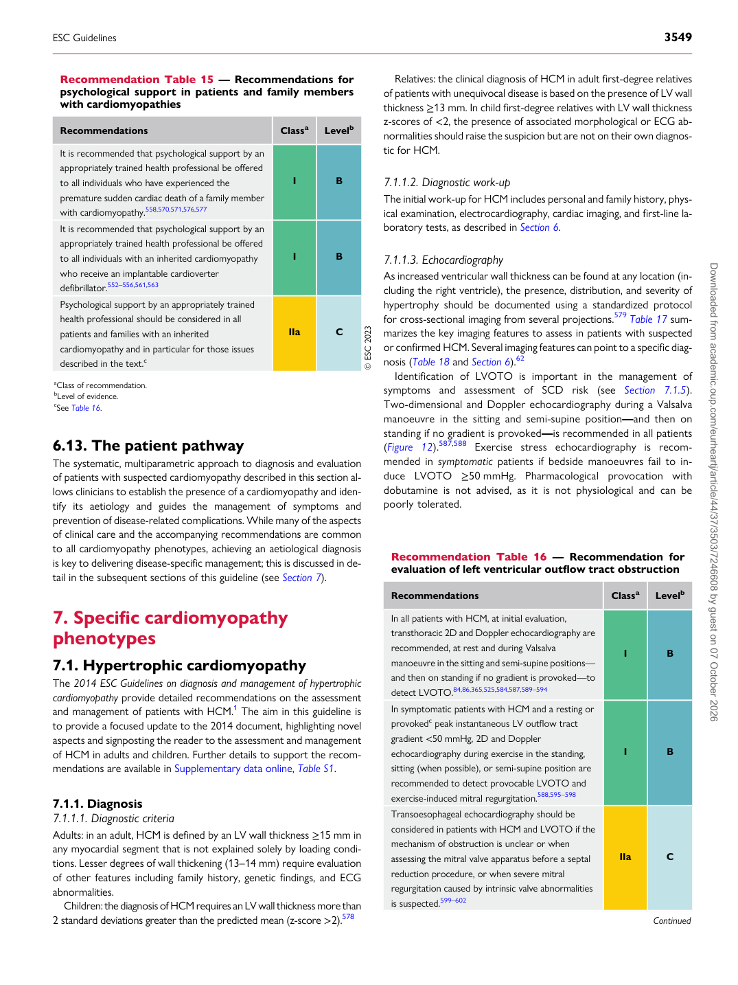
Recommendation Table 15 — Recommendations for psychological support in patients and family members with cardiomyopathies

Recommendations Classa Levelb

It is recommended that psychological support by an appropriately trained health professional be offered to all individuals who have experienced the premature sudden cardiac death of a family member with cardiomyopathy.558,570,571,576,577 I B It is recommended that psychological support by an appropriately trained health professional be offered to all individuals with an inherited cardiomyopathy who receive an implantable cardioverter defibrillator.552–556,561,563 I B Psychological support by an appropriately trained health professional should be considered in all patients and families with an inherited cardiomyopathy and in particular for those issues described in the text.c IIa C © ESC 2023 aClass of recommendation. bLevel of evidence. cSee Table 16.

Recommendation Table 16 — Recommendation for evaluation of left ventricular outflow tract obstruction

Recommendations Classa Levelb

In all patients with HCM, at initial evaluation, transthoracic 2D and Doppler echocardiography are recommended, at rest and during Valsalva manoeuvre in the sitting and semi-supine positions— and then on standing if no gradient is provoked—to detect LVOTO.84,86,365,525,584,587,589–594 I B In symptomatic patients with HCM and a resting or provokedc peak instantaneous LV outflow tract gradient <50 mmHg, 2D and Doppler echocardiography during exercise in the standing, sitting (when possible), or semi-supine position are recommended to detect provocable LVOTO and exercise-induced mitral regurgitation.588,595–598 I B Transoesophageal echocardiography should be considered in patients with HCM and LVOTO if the mechanism of obstruction is unclear or when assessing the mitral valve apparatus before a septal reduction procedure, or when severe mitral regurgitation caused by intrinsic valve abnormalities is suspected.599–602 IIa C

Continued

ESC Guidelines                                                                                                                                                                                          3549

Downloaded from academic.oup.com/eurheartj/article/44/37/3503/7246608 by guest on 07 October 2026