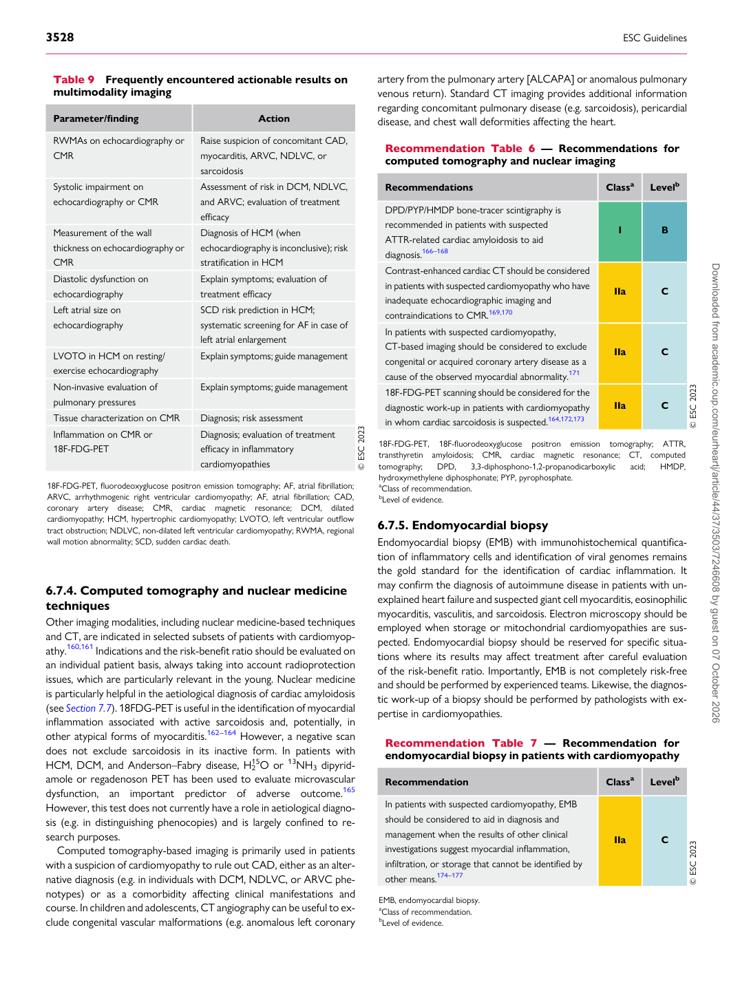
p.26 抽出テキスト
6.7.4 Computed tomography and nuclear medicine

techniques Other imaging modalities, including nuclear medicine-based techniques and CT, are indicated in selected subsets of patients with cardiomyopathy.160,161 Indications and the risk-benefit ratio should be evaluated on an individual patient basis, always taking into account radioprotection issues, which are particularly relevant in the young. Nuclear medicine is particularly helpful in the aetiological diagnosis of cardiac amyloidosis (see Section 7.7). 18FDG-PET is useful in the identification of myocardial inflammation associated with active sarcoidosis and, potentially, in other atypical forms of myocarditis.162–164 However, a negative scan does not exclude sarcoidosis in its inactive form. In patients with HCM, DCM, and Anderson–Fabry disease, H2

15O or 13NH3 dipyridamole or regadenoson PET has been used to evaluate microvascular dysfunction, an important predictor of adverse outcome.165 However, this test does not currently have a role in aetiological diagnosis (e.g. in distinguishing phenocopies) and is largely confined to research purposes. Computed tomography-based imaging is primarily used in patients with a suspicion of cardiomyopathy to rule out CAD, either as an alternative diagnosis (e.g. in individuals with DCM, NDLVC, or ARVC phenotypes) or as a comorbidity affecting clinical manifestations and course. In children and adolescents, CT angiography can be useful to exclude congenital vascular malformations (e.g. anomalous left coronary artery from the pulmonary artery [ALCAPA] or anomalous pulmonary venous return). Standard CT imaging provides additional information regarding concomitant pulmonary disease (e.g. sarcoidosis), pericardial disease, and chest wall deformities affecting the heart.

6.7.5 Endomyocardial biopsy

Endomyocardial biopsy (EMB) with immunohistochemical quantification of inflammatory cells and identification of viral genomes remains the gold standard for the identification of cardiac inflammation. It may confirm the diagnosis of autoimmune disease in patients with unexplained heart failure and suspected giant cell myocarditis, eosinophilic myocarditis, vasculitis, and sarcoidosis. Electron microscopy should be employed when storage or mitochondrial cardiomyopathies are suspected. Endomyocardial biopsy should be reserved for specific situations where its results may affect treatment after careful evaluation of the risk-benefit ratio. Importantly, EMB is not completely risk-free and should be performed by experienced teams. Likewise, the diagnostic work-up of a biopsy should be performed by pathologists with expertise in cardiomyopathies.

Table 9 Frequently encountered actionable results on multimodality imaging

Parameter/finding Action

RWMAs on echocardiography or CMR Raise suspicion of concomitant CAD,

myocarditis, ARVC, NDLVC, or sarcoidosis

Systolic impairment on echocardiography or CMR Assessment of risk in DCM, NDLVC, and ARVC; evaluation of treatment

efficacy

Measurement of the wall thickness on echocardiography or CMR Diagnosis of HCM (when

echocardiography is inconclusive); risk stratification in HCM

Diastolic dysfunction on echocardiography Explain symptoms; evaluation of

treatment efficacy

Left atrial size on echocardiography SCD risk prediction in HCM;

systematic screening for AF in case of left atrial enlargement

LVOTO in HCM on resting/ exercise echocardiography Explain symptoms; guide management Non-invasive evaluation of pulmonary pressures Explain symptoms; guide management Tissue characterization on CMR Diagnosis; risk assessment Inflammation on CMR or 18F-FDG-PET Diagnosis; evaluation of treatment

efficacy in inflammatory cardiomyopathies © ESC 2023

18F-FDG-PET, fluorodeoxyglucose positron emission tomography; AF, atrial fibrillation; ARVC, arrhythmogenic right ventricular cardiomyopathy; AF, atrial fibrillation; CAD, coronary artery disease; CMR, cardiac magnetic resonance; DCM, dilated cardiomyopathy; HCM, hypertrophic cardiomyopathy; LVOTO, left ventricular outflow tract obstruction; NDLVC, non-dilated left ventricular cardiomyopathy; RWMA, regional wall motion abnormality; SCD, sudden cardiac death. Recommendation Table 6 — Recommendations for computed tomography and nuclear imaging

Recommendations Classa Levelb

DPD/PYP/HMDP bone-tracer scintigraphy is recommended in patients with suspected ATTR-related cardiac amyloidosis to aid diagnosis.166–168 I B Contrast-enhanced cardiac CT should be considered in patients with suspected cardiomyopathy who have inadequate echocardiographic imaging and contraindications to CMR.169,170 IIa C In patients with suspected cardiomyopathy, CT-based imaging should be considered to exclude congenital or acquired coronary artery disease as a cause of the observed myocardial abnormality.171 IIa C 18F-FDG-PET scanning should be considered for the diagnostic work-up in patients with cardiomyopathy in whom cardiac sarcoidosis is suspected.164,172,173 IIa C © ESC 2023

18F-FDG-PET, 18F-fluorodeoxyglucose positron emission tomography; ATTR, transthyretin amyloidosis; CMR, cardiac magnetic resonance; CT, computed tomography; DPD, 3,3-diphosphono-1,2-propanodicarboxylic acid; HMDP, hydroxymethylene diphosphonate; PYP, pyrophosphate. aClass of recommendation. bLevel of evidence.

Recommendation Table 7 — Recommendation for endomyocardial biopsy in patients with cardiomyopathy

Recommendation Classa Levelb

In patients with suspected cardiomyopathy, EMB should be considered to aid in diagnosis and management when the results of other clinical investigations suggest myocardial inflammation, infiltration, or storage that cannot be identified by other means.174–177 IIa C © ESC 2023

EMB, endomyocardial biopsy. aClass of recommendation. bLevel of evidence. Downloaded from academic.oup.com/eurheartj/article/44/37/3503/7246608 by guest on 07 October 2026