p.25 抽出テキスト
Finding Cardiomyopathy

phenotype Cardiac CMR examples Specific diseases to be considered

Posterolateral LGE and concentric LVH Low native T1 Anderson–Fabry disease

Amyloidosis

Sarcomeric HCM

Haemochromatosis

Post-myocarditis

Dystrophinopathy

Sarcoidosis

Chagas disease

DSP variants FLNC variants DES variants

Laminopathy

Desmosomal variants

EMF/hypereosinophilia

Diffuse subendocardial LGE, high native T1

Patchy mid-wall in hypertrophied areas

Short T2*

Subepicardial LGE

Lateral wall epicardial LGE

Subepicardial and midwall LGE at basal septum +/- extension into inferolateral wall and RV insertion points

Apical transmural LGE

Ring-like and/or subepicardial LGE pattern

Septal mid-wall LGE

Fat and LGE (transmural RV plus sub-epicardial-midmural LV free wall)

Partial LV or RV apical obliteration + LGE at endocardial level

HCM

DCM

NDLVC

ARVC

RCM

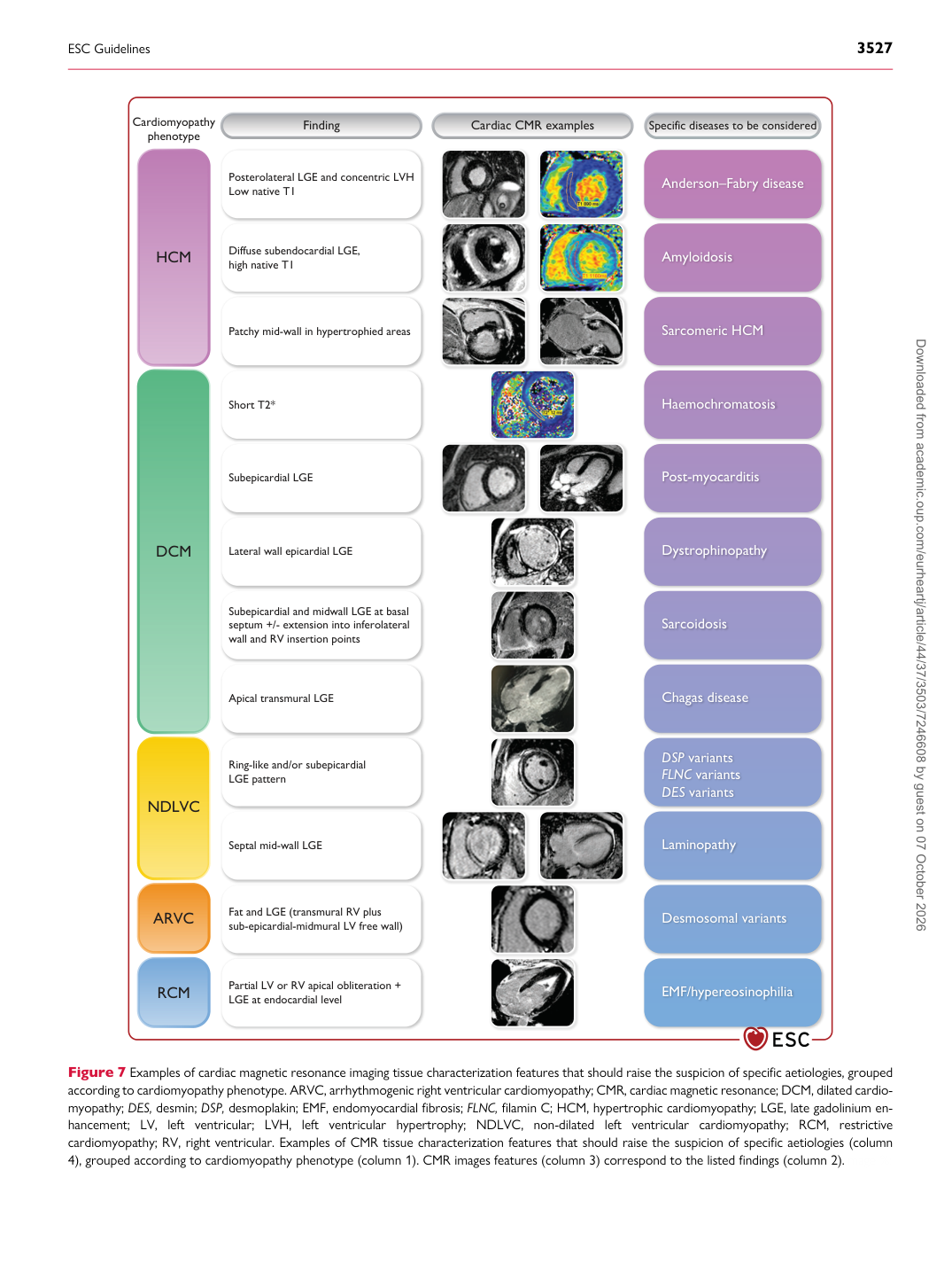
Figure 7 Examples of cardiac magnetic resonance imaging tissue characterization features that should raise the suspicion of specific aetiologies, grouped according to cardiomyopathy phenotype. ARVC, arrhythmogenic right ventricular cardiomyopathy; CMR, cardiac magnetic resonance; DCM, dilated cardiomyopathy; DES, desmin; DSP, desmoplakin; EMF, endomyocardial fibrosis; FLNC, filamin C; HCM, hypertrophic cardiomyopathy; LGE, late gadolinium enhancement; LV, left ventricular; LVH, left ventricular hypertrophy; NDLVC, non-dilated left ventricular cardiomyopathy; RCM, restrictive cardiomyopathy; RV, right ventricular. Examples of CMR tissue characterization features that should raise the suspicion of specific aetiologies (column 4), grouped according to cardiomyopathy phenotype (column 1). CMR images features (column 3) correspond to the listed findings (column 2).Image %:

ESC Guidelines                                                                                                                                                                                          3527

Downloaded from academic.oup.com/eurheartj/article/44/37/3503/7246608 by guest on 07 October 2026