p.23 抽出テキスト
6.7.3 Cardiac magnetic resonance

Cardiac magnetic resonance imaging (MRI) combines the advantages of non-invasiveness and independence of acoustic window with the ability for tissue characterization. The latter advantage is particularly important in the diagnosis of NDLVC, ARVC, myocarditis, amyloidosis, sarcoidosis and other forms of inflammatory disease, and iron overload/ haemochromatosis. Cardiac magnetic resonance is particularly useful if echocardiography provides poor image quality. Initial evaluation should routinely include cine imaging sequences, T2-weighted sequences, pre- and post-contrast T1 mapping, and late gadolinium enhancement (LGE). When suspecting haemochromatosis, T2* mapping should be employed. Cardiac magnetic resonance findings can provide important aetiological clues (Figure 7), with potential therapeutic implications (Table 9) and should be assessed collectively with genetic results and other clinical features by experienced operators in cardiac imaging and the evaluation of heart muscle disease. Serial follow-up CMR, every 2–5 years depending on initial severity and clinical course, can assist in evaluating disease progression as well as the benefits of therapy (e.g. evaluation of extracellular volume [ECV] in amyloidosis, or of iron deposition in haemochromatosis), and should be considered in all patients with cardiomyopathy.

6.7.3.1 Special considerations

• Recently developed rapid CMR techniques allow scans to be performed without general anaesthesia even in very young children.103 In children (and adults) unable to undergo CMR without general anaesthesia, the relative risks and benefits of the procedure should be considered. • Imaging artefacts caused by cardiac implantable electronic devices have posed limitations for CMR imaging in the past.104–110 A number of solutions are available to reduce artefacts, including reducing inhomogeneity, technical adjustments, and the use of special sequences, which reduce the rate of uninterpretable studies to one in five.111,112

Cardiac magnetic resonance can therefore be considered in patients with conditional devices and nearly all non-conditional devices provided appropriate protocols are put in place.113 • Nephrogenic systemic fibrosis is a rare complication reported in patients with first-generation linear unstable gadolinium chelates and

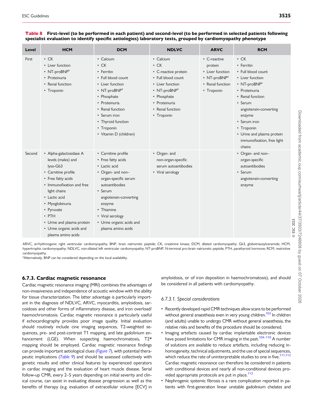
Table 8 First-level (to be performed in each patient) and second-level (to be performed in selected patients following specialist evaluation to identify specific aetiologies) laboratory tests, grouped by cardiomyopathy phenotype

Level HCM DCM NDLVC ARVC RCM

First • CK

• Liver function • NT-proBNPa • Proteinuria • Renal function • Troponin • Calcium

• CK • Ferritin • Full blood count • Liver function • NT-proBNPa • Phosphate • Proteinuria • Renal function • Serum iron • Thyroid function • Troponin • Vitamin D (children) • Calcium

• CK • C-reactive protein • Full blood count • Liver function • NT-proBNPa • Phosphate • Proteinuria • Renal function • Troponin • C-reactive

protein • Liver function • NT-proBNPa • Renal function • Troponin • CK

• Ferritin • Full blood count • Liver function • NT-proBNPa • Proteinuria • Renal function • Serum angiotensin-converting

enzyme • Serum iron • Troponin • Urine and plasma protein

immunofixation, free light chains

Second • Alpha-galactosidase A levels (males) and

lyso-Gb3 • Carnitine profile • Free fatty acids • Immunofixation and free light chains • Lactic acid • Myoglobinuria • Pyruvate • PTH • Urine and plasma protein • Urine organic acids and plasma amino acids • Carnitine profile • Free fatty acids

• Lactic acid • Organ- and non– organ-specific serum

autoantibodies • Serum angiotensin-converting

enzyme • Thiamine • Viral serology • Urine organic acids and

plasma amino acids • Organ- and non-organ-specific

serum autoantibodies • Viral serology • Organ- and non– organ-specific

autoantibodies • Serum angiotensin-converting

enzyme

© ESC 2023

ARVC, arrhythmogenic right ventricular cardiomyopathy; BNP, brain natriuretic peptide; CK, creatinine kinase; DCM, dilated cardiomyopathy; Gb3, globotriaosylceramide; HCM, hypertrophic cardiomyopathy; NDLVC, non-dilated left ventricular cardiomyopathy; NT-proBNP, N-terminal pro-brain natriuretic peptide; PTH, parathyroid hormone; RCM, restrictive cardiomyopathy. aAlternatively, BNP can be considered depending on the local availability.

ESC Guidelines                                                                                                                                                                                          3525

Downloaded from academic.oup.com/eurheartj/article/44/37/3503/7246608 by guest on 07 October 2026