p.22 抽出テキスト
6.6 Laboratory tests

Routine laboratory testing aids the detection of extracardiac conditions that cause or exacerbate ventricular dysfunction (e.g. thyroid disease, renal dysfunction, and diabetes mellitus) and secondary organ dysfunction in patients with severe heart failure. High levels of brain natriuretic peptide (BNP), N-terminal pro-brain natriuretic peptide (NT-proBNP), and high-sensitivity cardiac troponin T (hs-cTnT) are associated with cardiovascular events, heart failure, and death, and may have diagnostic, prognostic, and therapeutic monitoring value.69 Routine blood tests for comorbidities, including full blood count, renal and liver function parameters and electrolytes, thyroid function, fasting glucose, and Haemoglobin A1C (HbA1c) are recommended in all patients with heart failure symptoms.69 Persistently elevated serum creatinine kinase (CK) levels can be suggestive of myopathies or neuromuscular disorders including dystrophinopathies (e.g. Becker muscular dystrophy or X-linked DCM), laminopathies, desminopathies, or less often, a myofibrillar myopathy.62 Elevated C-reactive protein levels may be present in patients with ARVC and NDLVC, particularly in the context of recurrent myocarditis-like episodes.70 Elevated serum levels of iron and ferritin and high transferrin saturation can suggest a diagnosis of haemochromatosis and should trigger further aetiological refinement (primary vs. secondary) based on genetic testing. Lactic acidosis, myoglobinuria, and leucocytopaenia can be suggestive of mitochondrial diseases. A list of recommended laboratory tests in adults and children is shown in Table 8. Following specialist evaluation, additional tests to detect rare metabolic causes are often required in children, including measurement of lactate, pyruvate, pH, uric acid, ammonia, ketones, free fatty acids, carnitine profile, urine organic acids, and amino acids (see Section 6.9).

6.7 Multimodality imaging

6.7.1. General considerations Non-invasive imaging modalities represent the backbone of diagnosis and follow-up in patients with cardiomyopathies, including ultrasoundbased techniques, CMR imaging, computed tomography (CT), and nuclear techniques, such as positron emission tomography (PET) and scintigraphy (Figure 6).1,71,72 Physicians should always consider the yield of actionable results vs. the costs, advantages, and limitations of each technique, as well as patient safety and patient exposure to ionizing radiation and contrast media. Standardized algorithms should be in place to move hierarchically from simpler and cheaper to more complex and expensive tests. A bi-directional flow of information between the clinician and the imager is key to maximizing appropriateness: clinicians should formulate and share clear pre-test hypotheses, based on available information, to aid the interpretation of novel findings. The imager should respond in a similarly focused fashion, assessing the likelihood of alternative diagnoses and refraining from diagnoses that are not compatible/plausible based on the overall clinical context.

6.7.2 Echocardiography

The non-invasive nature and widespread availability of echocardiography make it the main imaging tool, from initial diagnosis to follow-up. Transthoracic echocardiography (TTE) provides relevant information on global and regional RV and LV anatomy and function as well as valve function and the presence of dynamic obstruction, pulmonary hypertension, or pericardial effusions.71–73 Myocardial deformation imaging (speckle tracking or tissue Doppler) with global longitudinal strain is a more sensitive marker than EF to detect subtle ventricular dysfunction (e.g. in genotype-positive HCM, DCM, and ARVC family members72,74,75), and may help discriminate between different aetiologies of hypertrophy76 (e.g. amyloidosis, HCM, and athlete’s heart). Mechanical dispersion is a marker of contraction inhomogeneity and highlights fine structural changes that may be missed by other modalities.77–80 Three-dimensional echocardiography reliably assesses volumes of cardiac chambers but needs an adequate acoustic window. Contrast agents can be considered for better endocardial delineation to depict the presence of hypertrabeculation, apical HCM, or apical aneurysms, and to exclude thrombus. Stress echocardiography can be helpful in selected patients to evaluate myocardial ischaemia and exercise echocardiography is useful to identify provocable LVOTO in symptomatic patients with HCM (see Section 7.1.1.3). Transoesophageal echocardiography is limited to selected indications, such as the exclusion of atrial thrombi related to AF, elucidating the mechanism of mitral regurgitation, or in planning invasive interventions (e.g. septal myectomy in HCM). When measuring cardiac dimensions and wall thickness in children, it is important to correct for body size, using z-scores (defined as the number of standard deviations from the population mean). Of note, there are inherent limitations with the use of z-scores in the diagnosis of cardiomyopathies, including the fact that there are many different normative data published resulting in significant variation in z-scores for the same patient.81 In addition, there are no normative data for wall thickness other than at the basal interventricular septum or posterior wall. The Task Force recommends using the normative data from the Paediatric Heart Network consortium.82

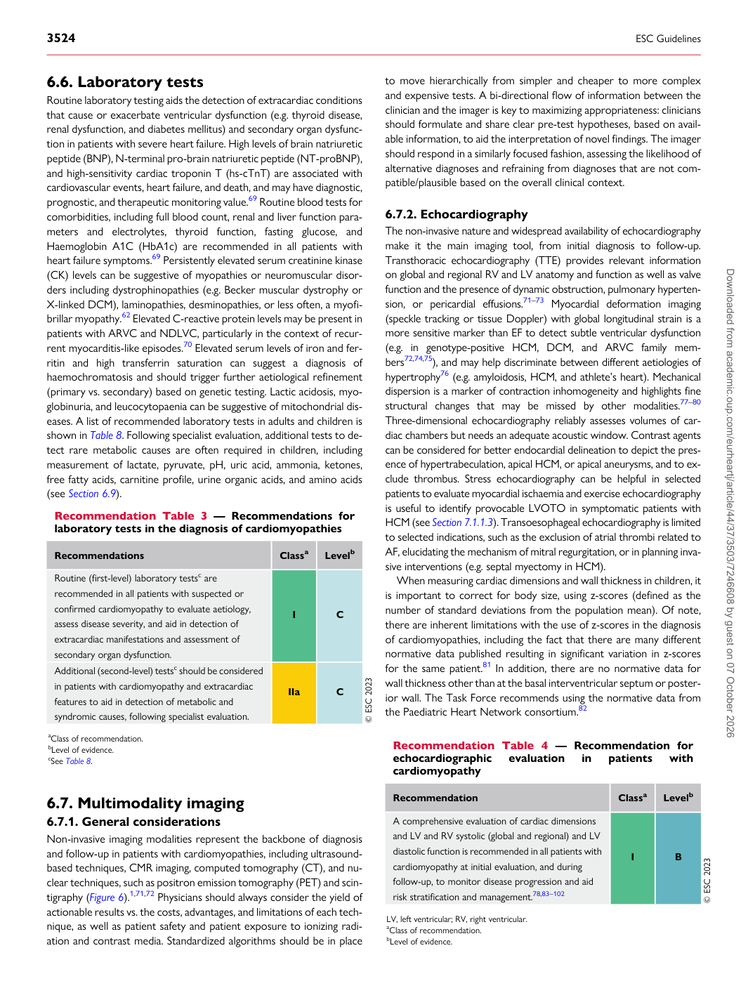
Recommendation Table 3 — Recommendations for laboratory tests in the diagnosis of cardiomyopathies

Recommendations Classa Levelb Routine (first-level) laboratory testsc are recommended in all patients with suspected or confirmed cardiomyopathy to evaluate aetiology, assess disease severity, and aid in detection of extracardiac manifestations and assessment of secondary organ dysfunction. I C Additional (second-level) testsc should be considered in patients with cardiomyopathy and extracardiac features to aid in detection of metabolic and syndromic causes, following specialist evaluation. IIa C © ESC 2023 aClass of recommendation. bLevel of evidence. cSee Table 8. Recommendation Table 4 — Recommendation for echocardiographic evaluation in patients with cardiomyopathy

Recommendation Classa Levelb

A comprehensive evaluation of cardiac dimensions and LV and RV systolic (global and regional) and LV diastolic function is recommended in all patients with cardiomyopathy at initial evaluation, and during follow-up, to monitor disease progression and aid risk stratification and management.78,83–102 I B © ESC 2023

LV, left ventricular; RV, right ventricular. aClass of recommendation. bLevel of evidence. Downloaded from academic.oup.com/eurheartj/article/44/37/3503/7246608 by guest on 07 October 2026