p.21 抽出テキスト
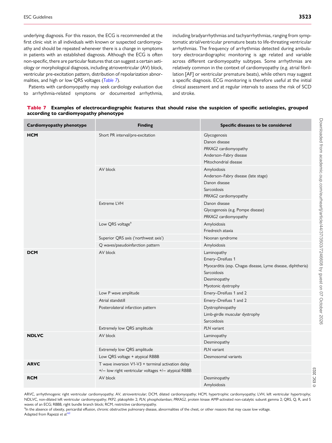
underlying diagnosis. For this reason, the ECG is recommended at the first clinic visit in all individuals with known or suspected cardiomyopathy and should be repeated whenever there is a change in symptoms in patients with an established diagnosis. Although the ECG is often non-specific, there are particular features that can suggest a certain aetiology or morphological diagnosis, including atrioventricular (AV) block, ventricular pre-excitation pattern, distribution of repolarization abnormalities, and high or low QRS voltages (Table 7). Patients with cardiomyopathy may seek cardiology evaluation due to arrhythmia-related symptoms or documented arrhythmia, including bradyarrhythmias and tachyarrhythmias, ranging from symptomatic atrial/ventricular premature beats to life-threating ventricular arrhythmias. The frequency of arrhythmias detected during ambulatory electrocardiographic monitoring is age related and variable across different cardiomyopathy subtypes. Some arrhythmias are relatively common in the context of cardiomyopathy (e.g. atrial fibrillation [AF] or ventricular premature beats), while others may suggest a specific diagnosis. ECG monitoring is therefore useful at the initial clinical assessment and at regular intervals to assess the risk of SCD and stroke.

Table 7 Examples of electrocardiographic features that should raise the suspicion of specific aetiologies, grouped according to cardiomyopathy phenotype

Cardiomyopathy phenotype Finding Specific diseases to be considered

HCM Short PR interval/pre-excitation Glycogenosis

Danon disease PRKAG2 cardiomyopathy Anderson–Fabry disease Mitochondrial disease

AV block Amyloidosis

Anderson–Fabry disease (late stage) Danon disease Sarcoidosis PRKAG2 cardiomyopathy

Extreme LVH Danon disease

Glycogenosis (e.g. Pompe disease) PRKAG2 cardiomyopathy

Low QRS voltagea Amyloidosis

Friedreich ataxia

Superior QRS axis (‘northwest axis’) Noonan syndrome Q waves/pseudoinfarction pattern Amyloidosis

DCM AV block Laminopathy Emery–Dreifuss 1

Myocarditis (esp. Chagas disease, Lyme disease, diphtheria) Sarcoidosis Desminopathy Myotonic dystrophy

Low P wave amplitude Emery–Dreifuss 1 and 2 Atrial standstill Emery–Dreifuss 1 and 2 Posterolateral infarction pattern Dystrophinopathy

Limb-girdle muscular dystrophy Sarcoidosis

Extremely low QRS amplitude PLN variant

NDLVC AV block Laminopathy Desminopathy

Extremely low QRS amplitude PLN variant Low QRS voltage + atypical RBBB Desmosomal variants

ARVC T wave inversion V1-V3 + terminal activation delay

+/− low right ventricular voltages +/− atypical RBBB

RCM AV block Desminopathy

Amyloidosis © ESC 2023

ARVC, arrhythmogenic right ventricular cardiomyopathy; AV, atrioventricular; DCM, dilated cardiomyopathy; HCM, hypertrophic cardiomyopathy; LVH, left ventricular hypertrophy; NDLVC, non-dilated left ventricular cardiomyopathy; PKP2, plakophilin 2; PLN, phospholamban; PRKAG2, protein kinase AMP-activated non-catalytic subunit gamma 2; QRS, Q, R, and S waves of an ECG; RBBB, right bundle branch block; RCM, restrictive cardiomyopathy. aIn the absence of obesity, pericardial effusion, chronic obstructive pulmonary disease, abnormalities of the chest, or other reasons that may cause low voltage. Adapted from Rapezzi et al.62

ESC Guidelines                                                                                                                                                                                          3523

Downloaded from academic.oup.com/eurheartj/article/44/37/3503/7246608 by guest on 07 October 2026