p.15 抽出テキスト
3.2.5 Restrictive cardiomyopathy

Restrictive cardiomyopathy (RCM) is defined as restrictive left and/or RV pathophysiology in the presence of normal or reduced diastolic volumes (of one or both ventricles), normal or reduced systolic volumes, and normal ventricular wall thickness.2 Restrictive cardiomyopathy commonly presents as biatrial enlargement. Left ventricular systolic function can be preserved, but it is rare for contractility to be completely normal. Restrictive pathophysiology may not be present throughout the natural history, but only at an initial stage (with an evolution towards a hypokinetic-dilated phase).11 Restrictive physiology can also occur in patients with end-stage hypertrophic and dilated cardiomyopathy; the preferred terms are ‘hypertrophic’ or ‘dilated cardiomyopathy with restrictive physiology’. Restrictive ventricular physiology can also be caused by endocardial pathology (fibrosis, fibroelastosis, and thrombosis) that impairs diastolic function.

3.3 Other traits and syndromes associated

with cardiomyopathy phenotypes 3.3.1. Left ventricular hypertrabeculation (left ventricular non-compaction) The term ‘left ventricular non-compaction’ (LVNC) has been used to describe a ventricular phenotype characterized by prominent LV trabeculae and deep intertrabecular recesses. The myocardial wall is often thickened with a thin, compacted epicardial layer and a thicker endocardial layer. In some patients, this abnormal trabecular architecture is associated with LV dilatation and systolic dysfunction. Left ventricular non-compaction is frequently a familial trait and is associated with variants in a range of genes, including those encoding proteins of the sarcomere, Z-disc, cytoskeleton, and nuclear envelope.12–16 Left ventricular non-compaction has also been used to describe an acquired and sometimes transient phenomenon of excessive LV trabeculation (e.g. in athletes, during pregnancy, or following vigorous activity)17–19 that must reflect increased prominence of an otherwise normal myocardial architecture, given that cardiomyocytes are terminally differentiated and the formation of new cardiac structures is impossible.20 The Task Force does not consider LVNC to be a cardiomyopathy in the general sense. Instead, it is seen as a phenotypic trait that can occur either in isolation or in association with other developmental abnormalities, ventricular hypertrophy, dilatation, and/or systolic dysfunction. Given the lack of morphometric evidence for ventricular compaction in humans,21,22 the term ‘hypertrabeculation’, rather than LVNC, is recommended, particularly when the phenomenon is transient or clearly of adult onset.

3.3.2 Takotsubo syndrome

Transient LV apical ballooning syndrome, or takotsubo syndrome, is characterized, in its most typical variant, by transient regional systolic dysfunction, dilatation, and oedema involving the LV apex and/or midventricle in the absence of obstructive coronary disease on coronary angiography.23 Patients present with an abrupt onset of angina-like chest pain and have diffuse T wave inversion (TWI), sometimes preceded by ST-segment elevation and mild cardiac enzyme elevation. Most reported cases occur in post-menopausal women. Symptoms are often preceded by emotional or physical stress. Norepinephrine concentration is elevated in most patients and a transient, dynamic outflow tract pressure gradient is reported in some cases. Left ventricular function usually normalizes over a period of days to weeks, and recurrence is rare. The same kind of reversible myocardial dysfunction is occasionally encountered in patients with intracranial haemorrhage or other acute cerebral accidents (neurogenic myocardial stunning).

Takotsubo syndrome is sometimes referred to as takotsubo or stress cardiomyopathy. Given the transient nature of the phenomenon, the Task Force does not recommend its classification as a cardiomyopathy.

4 Epidemiology

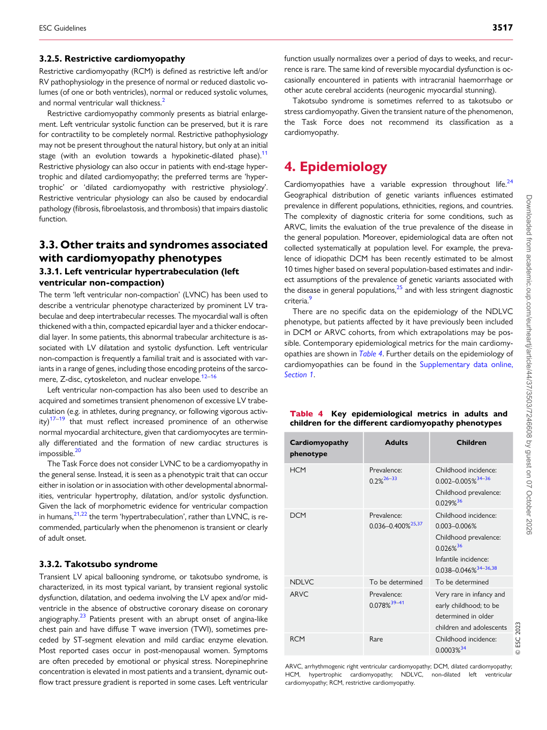
Cardiomyopathies have a variable expression throughout life.24 Geographical distribution of genetic variants influences estimated prevalence in different populations, ethnicities, regions, and countries. The complexity of diagnostic criteria for some conditions, such as ARVC, limits the evaluation of the true prevalence of the disease in the general population. Moreover, epidemiological data are often not collected systematically at population level. For example, the prevalence of idiopathic DCM has been recently estimated to be almost 10 times higher based on several population-based estimates and indirect assumptions of the prevalence of genetic variants associated with the disease in general populations,25 and with less stringent diagnostic criteria.9 There are no specific data on the epidemiology of the NDLVC phenotype, but patients affected by it have previously been included in DCM or ARVC cohorts, from which extrapolations may be possible. Contemporary epidemiological metrics for the main cardiomyopathies are shown in Table 4. Further details on the epidemiology of cardiomyopathies can be found in the Supplementary data online, Section 1.

Table 4 Key epidemiological metrics in adults and children for the different cardiomyopathy phenotypes

Cardiomyopathy phenotype Adults Children

HCM Prevalence:

0.2%26–33 Childhood incidence:

0.002–0.005%34–36 Childhood prevalence: 0.029%36

DCM Prevalence: 0.036–0.400%25,37 Childhood incidence: 0.003–0.006%

Childhood prevalence: 0.026%36 Infantile incidence: 0.038–0.046%34–36,38

NDLVC To be determined To be determined ARVC Prevalence: 0.078%39–41 Very rare in infancy and early childhood; to be

determined in older children and adolescents

RCM Rare Childhood incidence: 0.0003%34 © ESC 2023

ARVC, arrhythmogenic right ventricular cardiomyopathy; DCM, dilated cardiomyopathy; HCM, hypertrophic cardiomyopathy; NDLVC, non-dilated left ventricular cardiomyopathy; RCM, restrictive cardiomyopathy.

ESC Guidelines                                                                                                                                                                                          3517

Downloaded from academic.oup.com/eurheartj/article/44/37/3503/7246608 by guest on 07 October 2026