p.12 抽出テキスト
cardiomyopathy phenotypes may coexist in the same family, and that disease progression in an individual patient can include evolution from one cardiomyopathy phenotype to another. Nevertheless, the Task Force recommends an approach to disease nomenclature and diagnosis that is based on the predominant cardiac phenotype at presentation. While recognizing the fact that genes encoding cardiac ion channels may be implicated in some patients with dilated cardiomyopathy (DCM), conduction disorders, and arrhythmias, the Task Force was not persuaded that there is sufficient evidence to consider cardiac channelopathies as cardiomyopathies, in keeping with the approach taken by other recent ESC Guidelines.3 The most important changes in this guideline relate to the group of conditions variously included under the umbrella term ‘arrhythmogenic cardiomyopathies’. This term refers to a group of conditions that feature structural and functional abnormalities of the myocardium (identified by cardiac imaging and/or macroscopic and microscopic pathological investigation) and ventricular arrhythmia. This nosology has evolved in response to the recognition of the clinical and genetic overlap between right ventricular (RV) and LV cardiomyopathies, but a lack of a generally accepted definition has meant that the term encompasses a broad range of diverse pathologies and has introduced a number of inconsistencies and contradictions when applied in a clinical setting.4 The term ‘arrhythmogenic right ventricular (dysplasia/) cardiomyopathy’ (ARVC) was originally used by physicians who first discovered the disease, in the pre-genetic and pre-CMR era, to describe a new heart muscle disease predominantly affecting the right ventricle, whose cardinal clinical manifestation was the occurrence of malignant ventricular arrhythmias. Subsequently, autopsy investigations, genotype–phenotype correlation studies and the increasing use of contrast-enhancement CMR led to the identification of fibro-fatty replacement of the myocardium as a key phenotypic feature of the disease that affects the myocardium of both ventricles, with LV involvement which may even exceed the severity of RV involvement. This has led to the catch-all term of arrhythmogenic cardiomyopathy (ACM), which represents the evolution of the original term of ARVC.5 Consistent with its general approach, the Task Force agreed to highlight the vital importance of arrhythmia as a diagnostic red flag and prognostic marker across a range of clinical phenotypes, but did not recommend the use of the term ACM as a distinct cardiomyopathy subtype as it lacks a morphological or functional definition consistent with the existing classification scheme. While acknowledging that ‘ACM’ as an umbrella term that encompasses diverse clinical phenotypes has been previously used, this decision will, it is hoped, help to resolve many of the circular arguments that currently bedevil the field. The fundamental tenet throughout this guideline is that aetiology is vital to the management of patients with heart muscle disease and that a careful and consistent description of the morphological and functional phenotype is a crucial first step in the diagnostic pathway, while the final diagnosis will ideally describe aetiology alongside the phenotype.6,7

3.1 Definitions

A cardiomyopathy is defined as ‘a myocardial disorder in which the heart muscle is structurally and functionally abnormal, in the absence of coronary artery disease (CAD), hypertension, valvular disease, and congenital heart disease (CHD) sufficient to cause the observed myocardial abnormality’.2 This definition applies to both children and adults and makes no a priori assumptions about aetiology (which can be familial/genetic or acquired) or myocardial pathology. While the focus of this guideline is on genetic cardiomyopathies, the systematic approach to diagnosis starting from the phenotype at presentation described in this guideline enables clinicians to reach precise diagnoses that may also include non-genetic (e.g. inflammatory, toxic, and multisystem diseases) causes. It is important to note that cardiomyopathies can coexist with ischaemic, valvular, and hypertensive disease and that the presence of one does not exclude the possibility of the other.

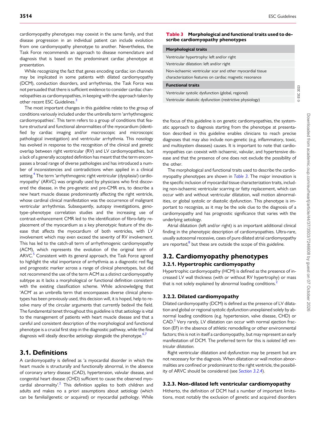
The morphological and functional traits used to describe the cardiomyopathy phenotypes are shown in Table 3. The major innovation is the specific inclusion of myocardial tissue characterization traits, including non-ischaemic ventricular scarring or fatty replacement, which can occur with and without ventricular dilatation, wall motion abnormalities, or global systolic or diastolic dysfunction. This phenotype is important to recognize, as it may be the sole clue to the diagnosis of a cardiomyopathy and has prognostic significance that varies with the underlying aetiology. Atrial dilatation (left and/or right) is an important additional clinical finding in the phenotypic description of cardiomyopathies. Ultra-rare, usually autosomal recessive, cases of pure dilated atrial cardiomyopathy are reported,8 but these are outside the scope of this guideline.

3.2 Cardiomyopathy phenotypes

3.2.1. Hypertrophic cardiomyopathy Hypertrophic cardiomyopathy (HCM) is defined as the presence of increased LV wall thickness (with or without RV hypertrophy) or mass that is not solely explained by abnormal loading conditions.2

3.2.2 Dilated cardiomyopathy

Dilated cardiomyopathy (DCM) is defined as the presence of LV dilatation and global or regional systolic dysfunction unexplained solely by abnormal loading conditions (e.g. hypertension, valve disease, CHD) or CAD.2 Very rarely, LV dilatation can occur with normal ejection fraction (EF) in the absence of athletic remodelling or other environmental factors; this is not in itself a cardiomyopathy, but may represent an early manifestation of DCM. The preferred term for this is isolated left ventricular dilatation. Right ventricular dilatation and dysfunction may be present but are not necessary for the diagnosis. When dilatation or wall motion abnormalities are confined or predominant to the right ventricle, the possibility of ARVC should be considered (see Section 3.2.4).

3.2.3 Non-dilated left ventricular cardiomyopathy

Hitherto, the definition of DCM had a number of important limitations, most notably the exclusion of genetic and acquired disorders

Table 3 Morphological and functional traits used to describe cardiomyopathy phenotypes Morphological traits Ventricular hypertrophy: left and/or right Ventricular dilatation: left and/or right Non-ischaemic ventricular scar and other myocardial tissue characterization features on cardiac magnetic resonance Functional traits Ventricular systolic dysfunction (global, regional) Ventricular diastolic dysfunction (restrictive physiology) © ESC 2023

Downloaded from academic.oup.com/eurheartj/article/44/37/3503/7246608 by guest on 07 October 2026