p.56 抽出テキスト
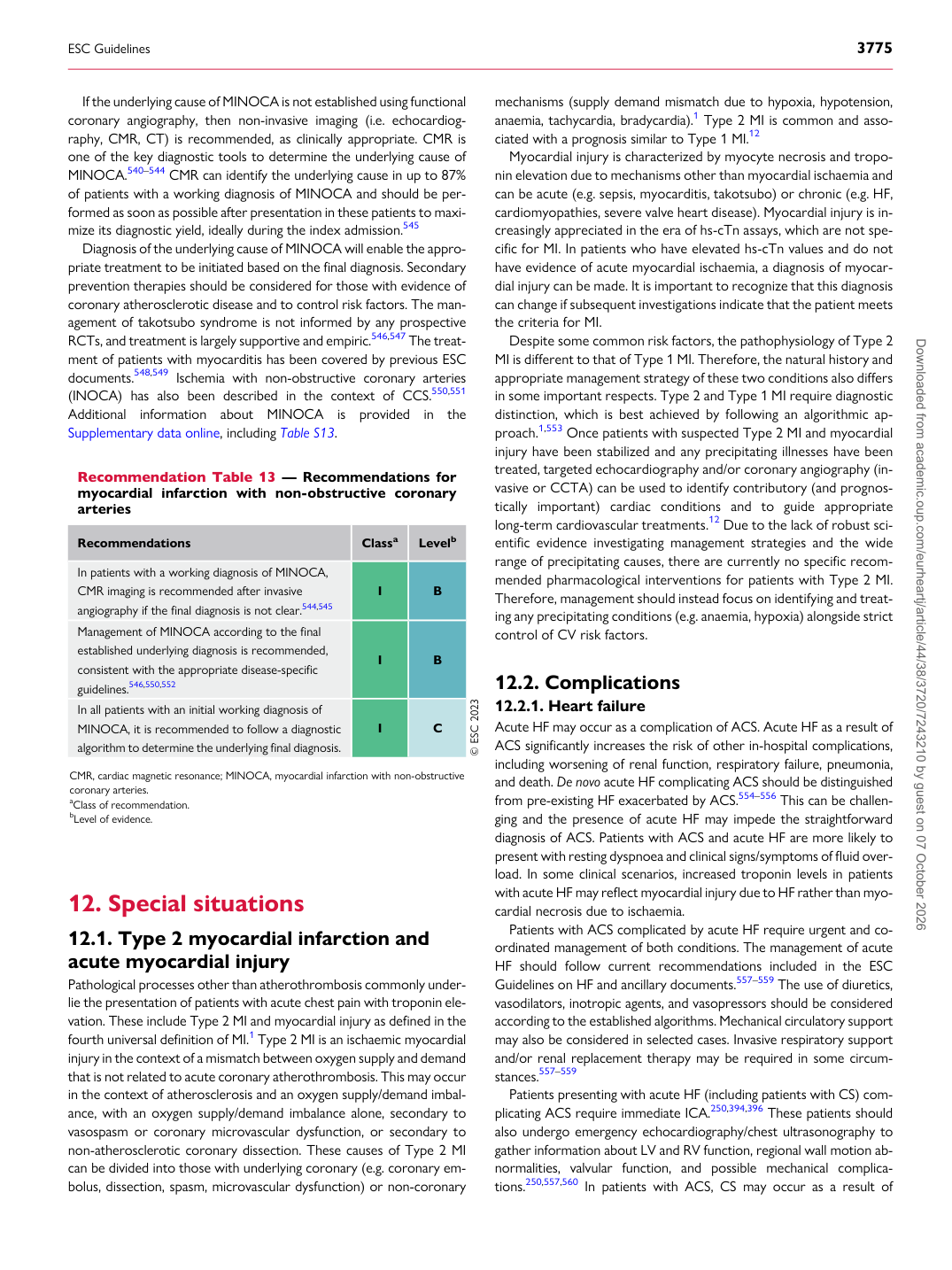
If the underlying cause of MINOCA is not established using functional coronary angiography, then non-invasive imaging (i.e. echocardiography, CMR, CT) is recommended, as clinically appropriate. CMR is one of the key diagnostic tools to determine the underlying cause of MINOCA.540–544 CMR can identify the underlying cause in up to 87% of patients with a working diagnosis of MINOCA and should be performed as soon as possible after presentation in these patients to maximize its diagnostic yield, ideally during the index admission.545 Diagnosis of the underlying cause of MINOCA will enable the appropriate treatment to be initiated based on the final diagnosis. Secondary prevention therapies should be considered for those with evidence of coronary atherosclerotic disease and to control risk factors. The management of takotsubo syndrome is not informed by any prospective RCTs, and treatment is largely supportive and empiric.546,547 The treatment of patients with myocarditis has been covered by previous ESC documents.548,549 Ischemia with non-obstructive coronary arteries (INOCA) has also been described in the context of CCS.550,551 Additional information about MINOCA is provided in the Supplementary data online, including Table S13.

12 Special situations

12.1 Type 2 myocardial infarction and

acute myocardial injury Pathological processes other than atherothrombosis commonly underlie the presentation of patients with acute chest pain with troponin elevation. These include Type 2 MI and myocardial injury as defined in the fourth universal definition of MI.1 Type 2 MI is an ischaemic myocardial injury in the context of a mismatch between oxygen supply and demand that is not related to acute coronary atherothrombosis. This may occur in the context of atherosclerosis and an oxygen supply/demand imbalance, with an oxygen supply/demand imbalance alone, secondary to vasospasm or coronary microvascular dysfunction, or secondary to non-atherosclerotic coronary dissection. These causes of Type 2 MI can be divided into those with underlying coronary (e.g. coronary embolus, dissection, spasm, microvascular dysfunction) or non-coronary mechanisms (supply demand mismatch due to hypoxia, hypotension, anaemia, tachycardia, bradycardia).1 Type 2 MI is common and associated with a prognosis similar to Type 1 MI.12

Myocardial injury is characterized by myocyte necrosis and troponin elevation due to mechanisms other than myocardial ischaemia and can be acute (e.g. sepsis, myocarditis, takotsubo) or chronic (e.g. HF, cardiomyopathies, severe valve heart disease). Myocardial injury is increasingly appreciated in the era of hs-cTn assays, which are not specific for MI. In patients who have elevated hs-cTn values and do not have evidence of acute myocardial ischaemia, a diagnosis of myocardial injury can be made. It is important to recognize that this diagnosis can change if subsequent investigations indicate that the patient meets the criteria for MI. Despite some common risk factors, the pathophysiology of Type 2 MI is different to that of Type 1 MI. Therefore, the natural history and appropriate management strategy of these two conditions also differs in some important respects. Type 2 and Type 1 MI require diagnostic distinction, which is best achieved by following an algorithmic approach.1,553 Once patients with suspected Type 2 MI and myocardial injury have been stabilized and any precipitating illnesses have been treated, targeted echocardiography and/or coronary angiography (invasive or CCTA) can be used to identify contributory (and prognostically important) cardiac conditions and to guide appropriate long-term cardiovascular treatments.12 Due to the lack of robust scientific evidence investigating management strategies and the wide range of precipitating causes, there are currently no specific recommended pharmacological interventions for patients with Type 2 MI. Therefore, management should instead focus on identifying and treating any precipitating conditions (e.g. anaemia, hypoxia) alongside strict control of CV risk factors.

12.2 Complications

12.2.1. Heart failure Acute HF may occur as a complication of ACS. Acute HF as a result of ACS significantly increases the risk of other in-hospital complications, including worsening of renal function, respiratory failure, pneumonia, and death. De novo acute HF complicating ACS should be distinguished from pre-existing HF exacerbated by ACS.554–556 This can be challenging and the presence of acute HF may impede the straightforward diagnosis of ACS. Patients with ACS and acute HF are more likely to present with resting dyspnoea and clinical signs/symptoms of fluid overload. In some clinical scenarios, increased troponin levels in patients with acute HF may reflect myocardial injury due to HF rather than myocardial necrosis due to ischaemia. Patients with ACS complicated by acute HF require urgent and coordinated management of both conditions. The management of acute HF should follow current recommendations included in the ESC Guidelines on HF and ancillary documents.557–559 The use of diuretics, vasodilators, inotropic agents, and vasopressors should be considered according to the established algorithms. Mechanical circulatory support may also be considered in selected cases. Invasive respiratory support and/or renal replacement therapy may be required in some circumstances.557–559 Patients presenting with acute HF (including patients with CS) complicating ACS require immediate ICA.250,394,396 These patients should also undergo emergency echocardiography/chest ultrasonography to gather information about LV and RV function, regional wall motion abnormalities, valvular function, and possible mechanical complications.250,557,560 In patients with ACS, CS may occur as a result of

Recommendation Table 13 — Recommendations for myocardial infarction with non-obstructive coronary arteries

Recommendations Classa Levelb

In patients with a working diagnosis of MINOCA, CMR imaging is recommended after invasive angiography if the final diagnosis is not clear.544,545 I B Management of MINOCA according to the final established underlying diagnosis is recommended, consistent with the appropriate disease-specific guidelines.546,550,552 I B In all patients with an initial working diagnosis of MINOCA, it is recommended to follow a diagnostic algorithm to determine the underlying final diagnosis. I C © ESC 2023

CMR, cardiac magnetic resonance; MINOCA, myocardial infarction with non-obstructive coronary arteries. aClass of recommendation. bLevel of evidence.

ESC Guidelines                                                                                                                                                                                          3775

Downloaded from academic.oup.com/eurheartj/article/44/38/3720/7243210 by guest on 07 October 2026