p.55 抽出テキスト
Cath lab assessment

Step 1

Clinical history Assessments to considera

Ward assessment

Step 2

Post discharge

care

Step 3 Detailed angiographic

assessment ± LV angiography (incl. LVeDP) Physical exam

Intravascular imaging (IVUS/OCT) ECG assessment

Assess for coronary microvascular dysfunction ± vasoreactivity (ACh testing)

Clinical history Assessments to considera

CMRI Physical exam

Blood testsb

ECG assessment

CTPA/CT brainc

Echocardiography

Assessments to considera

The MINOCA diagnostic algorithm

Follow-up clinic

evaluation Repeat echocardiography Repeat

CMRI Cardiac rehabilitation

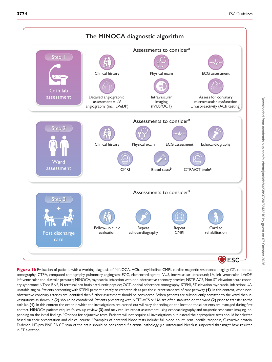
Figure 16 Evaluation of patients with a working diagnosis of MINOCA. ACh, acetylcholine; CMRI, cardiac magnetic resonance imaging; CT, computed tomography; CTPA, computed tomography pulmonary angiogram; ECG, electrocardiogram; IVUS, intravascular ultrasound; LV, left ventricular; LVeDP, left ventricular end-diastolic pressure; MINOCA, myocardial infarction with non-obstructive coronary arteries; NSTE-ACS, Non-ST elevation acute coronary syndrome; NTpro BNP, N terminal pro brain natriuretic peptide; OCT, optical coherence tomography; STEMI, ST-elevation myocardial infarction; UA, unstable angina. Patients presenting with STEMI present directly to catheter lab as per the current standard of care pathway (1). In this context, when nonobstructive coronary arteries are identified then further assessment should be considered. When patients are subsequently admitted to the ward then investigations as shown in (2) should be considered. Patients presenting with NSTE-ACS or UA are often stabilized on the ward (2) prior to transfer to the cath lab (1). In this context the order in which the investigations are carried out will vary depending on the location these patients are managed during first contact. MINOCA patients require follow-up review (3) and may require repeat assessment using echocardiography and magnetic resonance imaging, depending on the initial findings. aOptions for adjunctive tests. Patients will not require all investigations but instead the appropriate tests should be selected based on their presentation and clinical course. bExamples of potential blood tests include: full blood count, renal profile, troponin, C-reactive protein, D-dimer, NT-pro BNP. cA CT scan of the brain should be considered if a cranial pathology (i.e. intracranial bleed) is suspected that might have resulted in ST elevation. Downloaded from academic.oup.com/eurheartj/article/44/38/3720/7243210 by guest on 07 October 2026