p.48 抽出テキスト
9.1.2.2 Intravascular physiology

Intracoronary physiology is increasingly being used in patients with ACS to assess the haemodynamic significance of intermediate severity non-IRA stenoses (see Section 10). However, PCI of the IRA should not be deferred based on invasive epicardial functional assessment in patients with ACS. The coronary microcirculation begins to recover within 24 h of PPCI and acute functional assessment of the IRA may underestimate the true haemodynamic severity of the coronary stenosis.461 Beyond 1 week from the acute event, fractional flow reserve (FFR) measurement has been reported to reliably predict abnormal nuclear imaging results.462 Additional information about the role of intracoronary physiology in the IRA is presented in the Supplementary data online.

9.1.3 Timing of revascularization with percutaneous

coronary intervention In some patients with ACS undergoing ICA, an initial conservative management strategy with optimized guideline-directed medical therapy may be considered on a case-by-case basis. The specific circumstances include ACS patients with small calibre vessels, an occluded small side branch, or concerns regarding non-compliance with antithrombotic

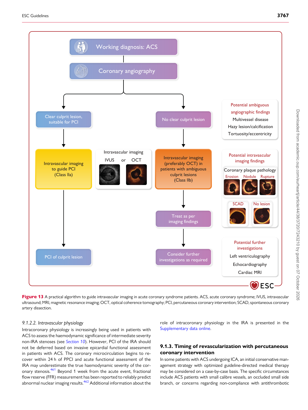
Working diagnosis: ACS

Clear culprit lesion,

suitable for PCI No clear culprit lesion

PCI of culprit lesion Consider further investigations as required

Treat as per imaging findings

Intravascular imaging

to guide PCI

(Class lla) Intravascular imaging Potential ambiguous

angiographic findings Multivessel disease Hazy lesion/calcification Tortuosity/eccentricity

Potential further

investigations Left ventriculography

Echocardiography

Cardiac MRI

Potential intravascular

imaging findings Coronary plaque pathology Erosion Nodule Rupture

IVUS or OCT Intravascular imaging

(preferably OCT) in patients with ambiguous

culprit lesions

(Class llb)

Coronary angiography

SCAD No lesion

Figure 13 A practical algorithm to guide intravascular imaging in acute coronary syndrome patients. ACS, acute coronary syndrome; IVUS, intravascular ultrasound; MRI, magnetic resonance imaging; OCT, optical coherence tomography; PCI, percutaneous coronary intervention; SCAD, spontaneous coronary artery dissection.

ESC Guidelines                                                                                                                                                                                          3767

Downloaded from academic.oup.com/eurheartj/article/44/38/3720/7243210 by guest on 07 October 2026