p.47 抽出テキスト
9 Technical aspects of invasive

strategies

9.1 Percutaneous coronary intervention

9.1.1. Vascular access Timely PCI with concomitant antithrombotic drugs has reduced the ischaemic risk in patients with ACS. However, this strategy is also associated with an increased bleeding risk, which affects prognosis at least as much as ischaemic complications and is associated with impaired survival.448,449 Among patients undergoing PCI, access-related bleeding accounts for 30–70% of total bleeding events.450 There is strong evidence demonstrating that reducing access-site bleeding events with the use of radial access translates into significant clinical benefits.448,449 The largest randomized trials on this topic in patients with ACS are the RadIal Vs femorAL access for coronary intervention (RIVAL) trial with 7021 ACS patients and the Minimizing Adverse Haemorrhagic Events by TRansradial Access Site and Systemic Implementation of angioX (MATRIX) trial with 8404 ACS patients (47.6% with STEMI).451,452 These trials have demonstrated significantly lower rates of access site-related bleeding, surgical access site repair, and blood transfusion with radial compared with femoral access. In the MATRIX trial, no significant interaction was observed between the type of ACS and the benefit associated with the radial approach, suggesting that the results of this trial can be extended to patients across the entire spectrum of ACS.453 In a costeffectiveness analysis of the MATRIX trial, radial access was also associated with significant savings in terms of quality-adjusted life years and PCI-related costs.454 Therefore, radial access is recommended as the preferred approach in ACS patients undergoing invasive assessment with or without PCI. However, femoral access may still be selectively chosen instead of radial access in certain patients (i.e. depending on the haemodynamic situation and other technical aspects during the index PCI procedure).

9.1.2 Intravascular imaging/physiology of the

infarct-related artery 9.1.2.1. Intravascular imaging As a diagnostic tool, intravascular imaging is useful in ACS patients without significant obstructive CAD on coronary angiography. Excluding an atherothrombotic cause in the main coronary arteries for the ACS may have important clinical implications, not only for immediate invasive management but also for potentially lifelong antithrombotic therapies. Intravascular imaging is also useful in cases where there is ambiguity regarding the culprit lesion. Culprit lesion ambiguity can be present in more than 30% of patients with suspected NSTE-ACS and over 10% of patients may have multiple culprit lesions.455,456 The recommendations for intravascular imaging in ACS are presented in Figure 13. The role of intravascular imaging is well established as a tool to guide and optimize PCI. Evidence in support of intravascular ultrasound (IVUS) guidance in ACS generally derives from subgroup analyses of allcomers trials. Meta-analysis of available randomized trials confirms the superiority of IVUS guidance in the reduction of MACE, although a definitive, large-scale, multinational trial is missing.457–459 Smaller RCTs have evaluated the role of optical coherence tomography (OCT) (see Supplementary data online).460

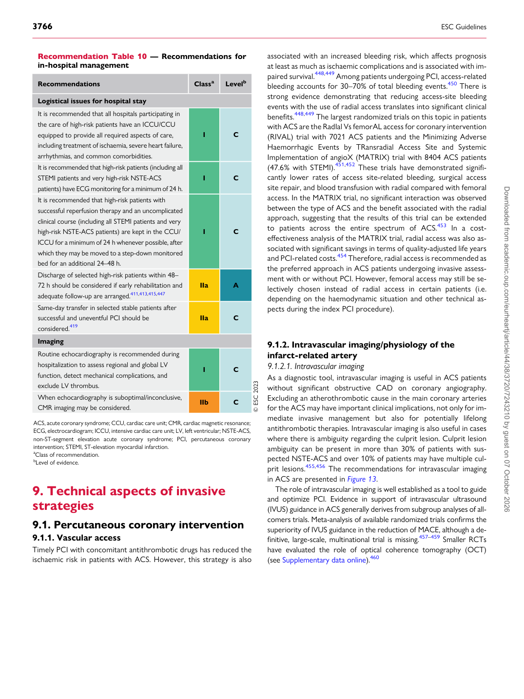
Recommendation Table 10 — Recommendations for in-hospital management

Recommendations Classa Levelb

Logistical issues for hospital stay It is recommended that all hospitals participating in the care of high-risk patients have an ICCU/CCU equipped to provide all required aspects of care, including treatment of ischaemia, severe heart failure, arrhythmias, and common comorbidities. I C It is recommended that high-risk patients (including all STEMI patients and very high-risk NSTE-ACS patients) have ECG monitoring for a minimum of 24 h. I C It is recommended that high-risk patients with successful reperfusion therapy and an uncomplicated clinical course (including all STEMI patients and very high-risk NSTE-ACS patients) are kept in the CCU/ ICCU for a minimum of 24 h whenever possible, after which they may be moved to a step-down monitored bed for an additional 24–48 h. I C Discharge of selected high-risk patients within 48– 72 h should be considered if early rehabilitation and adequate follow-up are arranged.411,413,415,447 IIa A Same-day transfer in selected stable patients after successful and uneventful PCI should be considered.419 IIa C Imaging Routine echocardiography is recommended during hospitalization to assess regional and global LV function, detect mechanical complications, and exclude LV thrombus. I C When echocardiography is suboptimal/inconclusive, CMR imaging may be considered. IIb C © ESC 2023

ACS, acute coronary syndrome; CCU, cardiac care unit; CMR, cardiac magnetic resonance; ECG, electrocardiogram; ICCU, intensive cardiac care unit; LV, left ventricular; NSTE-ACS, non-ST-segment elevation acute coronary syndrome; PCI, percutaneous coronary intervention; STEMI, ST-elevation myocardial infarction. aClass of recommendation. bLevel of evidence. Downloaded from academic.oup.com/eurheartj/article/44/38/3720/7243210 by guest on 07 October 2026