p.23 抽出テキスト
3.3.4.2 Practical guidance on how to implement the European

Society of Cardiology 0 h/1 h algorithm In order to maximize the safety and feasibility of implementing the 0 h/ 1 h algorithm, blood samples for hs-cTn at 0 h and 1 h should be obtained irrespective of other clinical details and pending results (see caveats of using rapid algorithms in Supplementary data online, Section 3.3. 2.2). This may result in unnecessary cTn measurements in the ∼10– 15% of patients with very low 0 h concentrations and chest pain onset >3 h, but substantially facilitates the process and thereby further increases patient safety. Similarly, the 0 h blood sample should be obtained immediately after admission to the ED.

3.3.5 Other biomarkers

The use of biomarkers other than cTn for the diagnosis of ACS is not recommended (unless cTn is not available). Among the multitude of additional biomarkers evaluated for the diagnosis of NSTEMI, only creatine kinase myocardial band isoenzyme, myosin-binding protein C, and copeptin may have clinical relevance when used in combination with (standard) cTn T/I, although in most clinical situations their incremental value above and beyond cTn is limited.45,46,83,102–114

3.4 Diagnostic tools | Non-invasive imaging

3.4.1. Echocardiography In emergency rooms and chest pain units, transthoracic echocardiography (TTE) performed or interpreted by trained healthcare professionals should be routinely available. In cases of suspected ACS with diagnostic uncertainty, TTE can be useful to identify signs suggestive of ongoing ischaemia or prior MI. However, this should not result in relevant delays in transfer to the cardiac catheterization laboratory if there is suspicion of an acute coronary artery occlusion. TTE can also be useful to suggest alternative aetiologies associated with chest pain (i.e. acute aortic disease, RV signs in pulmonary embolism [PE]). All patients presenting with CS or haemodynamic instability should undergo emergency TTE to try to identify the underlying cause—in particular, to assess LV and RV function and look for evidence of mechanical complications.

3.4.2 Computed tomography

Upon clinical presentation, CT is often the diagnostic tool of choice for ruling out alternative potentially life-threatening differential diagnoses of ACS, like PE or aortic dissection (this should be an ECG-gated contrast CT angiogram with full coverage of the thoracic aorta and the proximal head and neck vessels). Generally, CT does not have a role in patients presenting with suspicion of ongoing acute coronary occlusion, for whom emergency ICA is the priority. Coronary CT angiography (CCTA) has been investigated in many trials for the assessment of patients presenting to the ED with suspected NSTE-ACS. However, trials investigating CCTA in the era of hs-cTn assays may be of greater relevance for contemporary practice. The BEACON (Better Evaluation of Acute Chest Pain with Coronary Computed Tomography Angiography) study showed no reduction of inhospital duration of stay or hospital admission in the CCTA arm compared with patients investigated with hs-cTn, with similar results to those observed in the ROMICAT II (Rule Out Myocardial Ischemia/Infarction by Computer Assisted Tomography) and RAPID-CTCA (Rapid Assessment of Potential Ischaemic Heart Disease with CTCA) trials.115–117 In the latter study, a default approach using early non-invasive CCTA in patients with suspected NSTE-ACS did not improve clinical outcomes at 1 year and was associated with a modest increase in the duration and cost of the hospital stay. A default approach using CCTA as the first-line imaging investigation in patients with suspected NSTE-ACS is therefore not recommended. However, CCTA may provide added value in certain clinical settings (i.e. for patients in the observe zone in whom cTn and ECG results remain inconclusive). A normal CCTA (ruling out both obstructive and non-obstructive plaque) has a high NPV to exclude ACS and is associated with excellent clinical outcomes.

The systematic use of CCTA in rule-out patients after hospital discharge may identify the presence of obstructive or non-obstructive plaque and guide preventative medical therapies.118 CCTA can also be used to risk stratify selected low-risk NSTEMI patients. Such patients, who are found to have normal coronary arteries, non-obstructive coronary disease, or distal obstructive disease, may then not require ICA.119–121 Of note, the utility of CCTA may be limited in patients with tachycardia, established coronary artery disease (CAD), previous stents, or extensive coronary calcification.

3.4.3 Cardiac magnetic resonance imaging with or

without stress testing Cardiac magnetic resonance (CMR) imaging delineates cardiac structure and function, and also has the ability to provide assessments of myocardial perfusion and the pattern of myocardial injury. CMR is the imaging test of choice when poor echocardiographic windows preclude diagnostic echocardiographic evaluation. CMR allows direct visualization of infarcted regions, providing information on scarring and viability that can be differentiated from other forms of myocardial injury (e.g. myocarditis). CMR is therefore of particular clinical value in establishing a diagnosis of AMI where there is diagnostic uncertainty. CMR can also be useful in identifying the culprit vascular territory and in confirming a diagnosis of myocarditis or takotsubo cardiomyopathy, amongst other differentials. CMR is of particular value in establishing a diagnosis in patients presenting with a working diagnosis of myocardial infarction with non-obstructive coronary arteries (MINOCA) following invasive angiography and is the gold standard for the assessment of LV thrombus.

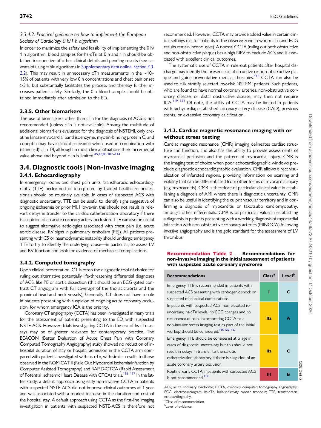
Recommendation Table 2 — Recommendations for non-invasive imaging in the initial assessment of patients with suspected acute coronary syndrome

Recommendations Classa Levelb

Emergency TTE is recommended in patients with suspected ACS presenting with cardiogenic shock or suspected mechanical complications. I C In patients with suspected ACS, non-elevated (or uncertain) hs-cTn levels, no ECG changes and no recurrence of pain, incorporating CCTA or a non-invasive stress imaging test as part of the initial workup should be considered.116,122–127 IIa A Emergency TTE should be considered at triage in cases of diagnostic uncertainty but this should not result in delays in transfer to the cardiac catheterization laboratory if there is suspicion of an acute coronary artery occlusion. IIa C Routine, early CCTA in patients with suspected ACS is not recommended.117 III B © ESC 2023

ACS, acute coronary syndrome; CCTA, coronary computed tomography angiography; ECG, electrocardiogram; hs-cTn, high-sensitivity cardiac troponin; TTE, transthoracic echocardiography. aClass of recommendation. bLevel of evidence. Downloaded from academic.oup.com/eurheartj/article/44/38/3720/7243210 by guest on 07 October 2026