p.19 抽出テキスト
elevation (or ST-segment elevation equivalents) on ECG (working diagnosis: non-ST-elevation [NSTE]-ACS). These patients may exhibit other ECG alterations, including transient ST-segment elevation, persistent or transient ST-segment depression, and T wave abnormalities, including hyperacute T waves, T wave inversion, biphasic T waves, flat T waves, and pseudonormalization of T waves. Alternatively, the ECG may be normal. The majority of patients in this category who subsequently display a typical rise and fall in cardiac troponin levels (i.e. fulfilling MI criteria as per the fourth universal definition of MI) will receive a final diagnosis of non-ST-elevation MI (NSTEMI). In other patients, the troponin level will remain below the 99th centile and they will receive a final diagnosis of UA, although with high-sensitivity troponin assays this diagnosis has become less common. It is also important to recognize that NSTEMI or UA will not be the final diagnosis in all patients with an initial working diagnosis of NSTE-ACS.

3.2.1 Acute coronary syndrome with persistent

ST-segment elevation (suspected ST-elevation myocardial infarction) The priority for these patients is the implementation of reperfusion therapy as soon as possible (see Section 5). In the appropriate clinical context, ST-segment elevation (measured at the J-point) is considered suggestive of ongoing coronary artery acute occlusion in the following cases:

New ST elevation at the J-point in at least two contiguous leads: • ≥2.5 mm in men <40 years, ≥2 mm in men ≥40 years, or ≥1.5 mm in women regardless of age in leads V2–V3 • and/or ≥1 mm in the other leads (in the absence of left ventricular [LV] hypertrophy or left bundle branch block [LBBB]).

In patients with suspected inferior STEMI, it is recommended to record right precordial leads (V3R and V4R) in order to assess for ST-segment elevation.6 Posterior leads (V7–V9) can also be recorded to investigate for posterior STEMI, particularly in patients with ongoing symptoms and an inconclusive standard 12-lead ECG. The diagnosis of ongoing acute coronary artery occlusion on ECG can sometimes be challenging, and some cases may warrant prompt management and triage for immediate reperfusion therapy despite the absence of ST-segment elevation. It is also important to recognize that while the most sensitive sign for ongoing acute coronary artery occlusion is ST-segment elevation, there are other ECG findings that can be suggestive of ongoing coronary artery occlusion (or severe ischaemia). If these findings are present, prompt triage for immediate reperfusion therapy is indicated (see Supplementary data online, Figure S2). ST-segment depression in leads V1–V3 (especially when the terminal T wave is positive) and/or ST-segment elevation in V7–V9 are highly suggestive of posterior coronary artery occlusion (often the left circumflex artery).1,7 ST-segment elevation in V3R and V4R is highly suggestive of ongoing RV ischaemia.8 ST depression ≥1 mm in ≥6 surface leads (inferolateral ST depression), coupled with ST-segment elevation in aVR and/or V1, suggests multivessel ischaemia or left main coronary artery obstruction, particularly if the patient presents with haemodynamic compromise.9–11

Bundle branch block (BBB). In patients with a high clinical suspicion of ongoing myocardial ischaemia, the presence of LBBB, right bundle branch block (RBBB), or a paced rhythm precludes an accurate assessment of the presence or absence of ST-segment elevation. Therefore, patients presenting with these ECG patterns in combination with signs/symptoms that are highly suspicious for ongoing myocardial ischaemia should be managed similarly to those with clear ST-segment elevation, regardless of whether the BBB is previously known (see Supplementary data online).4

3.2.2 Acute coronary syndrome without persistent

ST-segment elevation (non-ST elevation acute coronary syndrome) While the ECG in the setting of NSTE-ACS may be normal in more than one-third of patients, characteristic ECG abnormalities are frequently present and increase the diagnostic probability of ACS.12–16 These ECG abnormalities include ST depression and T wave changes (especially biphasic T waves or prominent negative T waves [Wellens’ sign, related to severe proximal left anterior descending artery stenosis]), (see Supplementary data online, Figure S3).

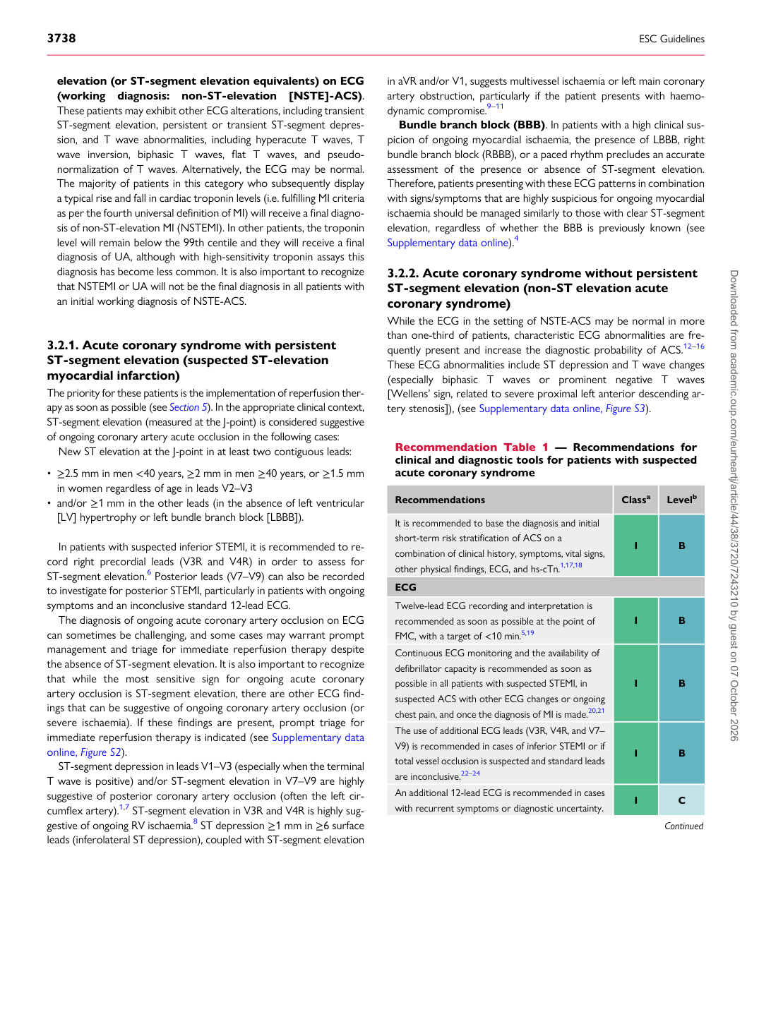
Recommendation Table 1 — Recommendations for clinical and diagnostic tools for patients with suspected acute coronary syndrome

Recommendations Classa Levelb

It is recommended to base the diagnosis and initial short-term risk stratification of ACS on a combination of clinical history, symptoms, vital signs, other physical findings, ECG, and hs-cTn.1,17,18 I B ECG Twelve-lead ECG recording and interpretation is recommended as soon as possible at the point of FMC, with a target of <10 min.5,19 I B Continuous ECG monitoring and the availability of defibrillator capacity is recommended as soon as possible in all patients with suspected STEMI, in suspected ACS with other ECG changes or ongoing chest pain, and once the diagnosis of MI is made.20,21 I B The use of additional ECG leads (V3R, V4R, and V7– V9) is recommended in cases of inferior STEMI or if total vessel occlusion is suspected and standard leads are inconclusive.22–24 I B An additional 12-lead ECG is recommended in cases with recurrent symptoms or diagnostic uncertainty. I C

Continued Downloaded from academic.oup.com/eurheartj/article/44/38/3720/7243210 by guest on 07 October 2026