p.12 抽出テキスト
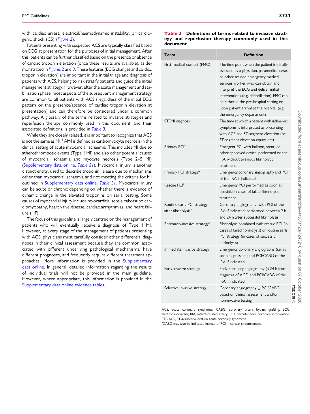
with cardiac arrest, electrical/haemodynamic instability, or cardiogenic shock (CS) (Figure 2). Patients presenting with suspected ACS are typically classified based on ECG at presentation for the purposes of initial management. After this, patients can be further classified based on the presence or absence of cardiac troponin elevation (once these results are available), as demonstrated in Figures 2 and 3. These features (ECG changes and cardiac troponin elevation) are important in the initial triage and diagnosis of patients with ACS, helping to risk stratify patients and guide the initial management strategy. However, after the acute management and stabilization phase, most aspects of the subsequent management strategy are common to all patients with ACS (regardless of the initial ECG pattern or the presence/absence of cardiac troponin elevation at presentation) and can therefore be considered under a common pathway. A glossary of the terms related to invasive strategies and reperfusion therapy commonly used in this document, and their associated definitions, is provided in Table 3. While they are closely related, it is important to recognize that ACS is not the same as MI.1 AMI is defined as cardiomyocyte necrosis in the clinical setting of acute myocardial ischaemia. This includes MI due to atherothrombotic events (Type 1 MI) and also other potential causes of myocardial ischaemia and myocyte necrosis (Type 2–5 MI) (Supplementary data online, Table S1). Myocardial injury is another distinct entity, used to describe troponin release due to mechanisms other than myocardial ischaemia and not meeting the criteria for MI outlined in Supplementary data online, Table S1. Myocardial injury can be acute or chronic depending on whether there is evidence of dynamic change in the elevated troponins on serial testing. Some causes of myocardial injury include myocarditis, sepsis, takotsubo cardiomyopathy, heart valve disease, cardiac arrhythmias, and heart failure (HF). The focus of this guideline is largely centred on the management of patients who will eventually receive a diagnosis of Type 1 MI. However, at every stage of the management of patients presenting with ACS, physicians must carefully consider other differential diagnoses in their clinical assessment because they are common, associated with different underlying pathological mechanisms, have different prognoses, and frequently require different treatment approaches. More information is provided in the Supplementary data online. In general, detailed information regarding the results of individual trials will not be provided in the main guideline. However, where appropriate, this information is provided in the Supplementary data online evidence tables. Table 3 Definitions of terms related to invasive strategy and reperfusion therapy commonly used in this document

Term Definition

First medical contact (FMC) The time point when the patient is initially assessed by a physician, paramedic, nurse,

or other trained emergency medical services worker who can obtain and interpret the ECG and deliver initial interventions (e.g. defibrillation). FMC can be either in the pre-hospital setting or upon patient arrival at the hospital (e.g. the emergency department)

STEMI diagnosis The time at which a patient with ischaemic

symptoms is interpreted as presenting with ACS and ST-segment elevation (or ST-segment elevation equivalent)

Primary PCIa Emergent PCI with balloon, stent, or

other approved device, performed on the IRA without previous fibrinolytic treatment

Primary PCI strategya Emergency coronary angiography and PCI of the IRA if indicated Rescue PCIa Emergency PCI performed as soon as possible in cases of failed fibrinolytic

treatment

Routine early PCI strategy after fibrinolysisa Coronary angiography, with PCI of the

IRA if indicated, performed between 2 h and 24 h after successful fibrinolysis

Pharmaco-invasive strategya Fibrinolysis combined with rescue PCI (in

cases of failed fibrinolysis) or routine early PCI strategy (in cases of successful fibrinolysis)

Immediate invasive strategy Emergency coronary angiography (i.e. as

soon as possible) and PCI/CABG of the IRA if indicated

Early invasive strategy Early coronary angiography (<24 h from

diagnosis of ACS) and PCI/CABG of the IRA if indicated

Selective invasive strategy Coronary angiography ± PCI/CABG based on clinical assessment and/or

non-invasive testing © ESC 2023

ACS, acute coronary syndrome; CABG, coronary artery bypass grafting; ECG, electrocardiogram; IRA, infarct-related artery; PCI, percutaneous coronary intervention; STE-ACS, ST-segment-elevation acute coronary syndrome. aCABG may also be indicated instead of PCI in certain circumstances.

ESC Guidelines                                                                                                                                                                                          3731

Downloaded from academic.oup.com/eurheartj/article/44/38/3720/7243210 by guest on 07 October 2026