p.83 抽出テキスト
7.2.3. Andersen–Tawil syndrome Type 1 Andersen–Tawil syndrome Type 1, also classified as LQT7, is a rare disease (1:1 000000) characterized by three main symptoms: frequent VA (e.g. bidirectional VT), dysmorphologies and periodic paralysis.964–967 The inward rectifier current (IK1) decreased by KCNJ2 loss of function mutation968 causes an increase in U wave amplitude rather than QT prolongation.964,967–970 Syncope or documented VT is associated with life-threatening VA, and a study found a 5-year probability of 7.9% of life-threatening VA.967 In patients with sustained haemodynamically not-tolerated VT or CA, an ICD is recommended.964,967 Flecainide and/or beta-blockers seem to reduce VA,964,970,971 whereas amiodarone may be proarrhythmic and should only be used with an ICD.967 In patients with syncope despite medical therapy, an ICD or ILR should be discussed.967 7.2.4 Brugada syndrome

The type 1 Brugada ECG pattern is characterized by J point elevation of .2 mV with coved ST elevation and T wave inversion in at least one right precordial ECG lead, V1 or V2, positioned in the second, third or fourth intercostal spaces (Figure 32). It may occur either spontaneously or be induced by exposure to sodium channelblocking drugs or fever.135,231,973–978 It is mandatory to exclude other conditions that may explain the type 1 pattern, so-called phenocopies.979 BrS is diagnosed in patients without other heart disease and a spontaneous type 1 pattern, regardless of symptoms, due to its rarity in the general population and association with risk.231,979,980 The type 1 pattern may also be induced by administration of a sodium channel-blocking drug as a diagnostic test in patients suspected to have concealed BrS but without a spontaneous type 1 ECG.135,136,231,387,973,978,981–985 However, provocation with drug or fever is less specific than previously thought, with 2–4% prevalence in healthy subjects and higher prevalence in patients with AV nodal reentry tachycardia or an accessory pathway in one study.977,978,986 In the opinion of this panel of experts, an induced type 1 ECG pattern therefore requires other clinical features, such as documented PVT/VF, arrhythmic syncope, or relevant family history. The yield of genetic testing in BrS patients is approximately 20%, with the SCN5A gene the only gene with evidence of association for clinical testing purposes.164,980 Phenotype and genotype mismatch is seen in SCN5A families, which is explained by variable effects of mutation severity and a polygenic risk score derived from genome-wide association studies.170,979,987 Recent data also support a potential for prognostication.988,989 Psychotropic drugs, selected AADs, anaesthetic agents, cocaine, excessive alcohol intake and fever are potential triggers to exacerbate the type 1 pattern and trigger VF.231,297 The risk of recurrent VF among patients presenting with CA is 48% at 10 years. ICD implantation is therefore indicated in symptomatic BrS patients who are survivors of CA or have documented spontaneous sustained VA (Figure 33).980,990–994 Approximately one-third of BrS patients present with syncope.995 The risk of arrhythmic events in BrS patients with unexplained syncope is 4 times higher than the risk in asymptomatic patients.155,990–992,994,996 Detailed patient history, including absence of prodrome or specific triggers, is essential for distinguishing arrhythmic from non-arrhythmic syncope. Nonetheless, the aetiology of syncope is difficult to determine in up to 30% of BrS patients. In small studies, arrhythmia detected by ILR changed clinical management in 20–36% of the BrS patients with unexplained syncope.997–999 Asymptomatic patients represent a majority of newly diagnosed BrS patients with an incidence of arrhythmic events of 0.5% per year.1000,1001 Their risk stratification remains challenging. A spontaneous type 1 ECG pattern, as well as other ECG markers such as early repolarization pattern and QRS fragmentation, have been associated with higher risk.980,992,1002,1003 Some have been incorporated into risk scores, although their utility in intermediate-risk patients remains low.1004,1005 Electrophysiological studies remain controversial. A multicentre pooled analysis showed that

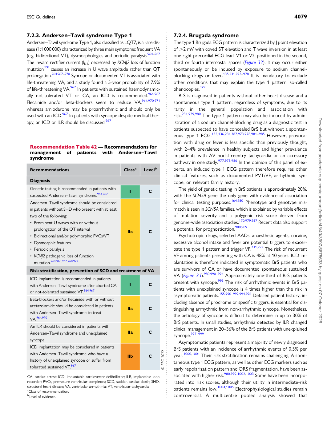
Recommendation Table 42 — Recommendations for management of patients with Andersen–Tawil syndrome

Recommendations Classa Levelb

Diagnosis Genetic testing is recommended in patients with suspected Andersen–Tawil syndrome.964,967 I C Andersen–Tawil syndrome should be considered in patients without SHD who present with at least two of the following: • Prominent U waves with or without

prolongation of the QT interval • Bidirectional and/or polymorphic PVCs/VT • Dysmorphic features • Periodic paralysis • KCNJ2 pathogenic loss of function mutation.964,965,967,968,972 IIa C Risk stratification, prevention of SCD and treatment of VA ICD implantation is recommended in patients with Andersen–Tawil syndrome after aborted CA or not-tolerated sustained VT.964,967 I C Beta-blockers and/or flecainide with or without acetazolamide should be considered in patients with Andersen–Tawil syndrome to treat VA.964,970 IIa C An ILR should be considered in patients with Andersen–Tawil syndrome and unexplained syncope. IIa C ICD implantation may be considered in patients with Andersen–Tawil syndrome who have a history of unexplained syncope or suffer from tolerated sustained VT.967 IIb C © ESC 2022

CA, cardiac arrest; ICD, implantable cardioverter defibrillator; ILR, implantable loop recorder; PVCs, premature ventricular complexes; SCD, sudden cardiac death; SHD, structural heart disease; VA, ventricular arrhythmia; VT, ventricular tachycardia. aClass of recommendation. bLevel of evidence. Downloaded from academic.oup.com/eurheartj/article/43/40/3997/6675633 by guest on 07 October 2026