p.63 抽出テキスト
patients with severe RV dysfunction (RV fractional area change ≤17% or RV ejection fraction ≤35%), an ICD should be considered. Likewise, ARVC patients with significant LV involvement (LVEF ≤35%) should be treated according to the current DCM recommendation regarding ICD implantation. There are conflicting data on the independent predictive value of NSVT for subsequent sustained arrhythmic events.695,696,701 Likewise, the role of PES in risk stratification, particularly in asymptomatic patients, is not well defined.695,696,705 In one series including mainly symptomatic patients, both NSVT and a positive PES independently predicted arrhythmic events, while others report a low diagnostic accuracy.696 Risk prediction based on a single parameter does not consider the potential combined effect and interactions between factors. Therefore, although supporting data are lacking, this panel of experts supports that an ICD should be considered706 in symptomatic ARVC patients with moderate RV (,40%) and/or LV dysfunction (,45%) and who have either NSVT or have SMVT inducible at PES. An internally validated risk model has recently been developed from 528 patients with definite ARVC and no prior VA, including age, sex, arrhythmic syncope, NSVT, PVC burden, leads with T-wave inversion and RV ejection fraction, to predict any sustained VA (c-index 0.77).81 In addition, a prediction model for specifically life-threatening VA has been promoted based on 864 ARVC patients, with predictors including male sex, age, 24 h PVC count, and leads with T-wave inversion (c-index 0.74).702 However, validation studies are needed before these risk models can be recommended for clinical use.

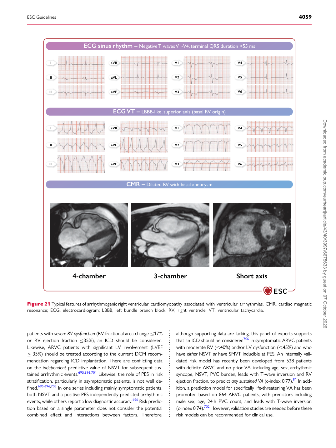
ECG sinus rhythm – Negative T wavesV1-V4, terminal QRS duration >55 ms

ECG VT – LBBB-like, superior axis (basal RV origin)

CMR – Dilated RV with basal aneurysm

4-chamber 3-chamber Short axis

I

II

III V1

V2

V3

aVR

aVL

aVF V4

V5

V6

I

II

III V1

V2

V3

aVR

aVL

aVF V4

V5

V6

Figure 21 Typical features of arrhythmogenic right ventricular cardiomyopathy associated with ventricular arrhythmias. CMR, cardiac magnetic resonance; ECG, electrocardiogram; LBBB, left bundle branch block; RV, right ventricle; VT, ventricular tachycardia. Downloaded from academic.oup.com/eurheartj/article/43/40/3997/6675633 by guest on 07 October 2026